Mass: 18.015 Da / Num. of mol.: 162 / Source method: isolated from a natural source / Formula: H2O
Compound details
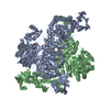
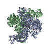
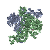
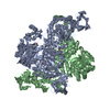
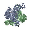
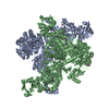
ALPHA-L-FUCOSIDASE BELONGS TO GLYCOSYL HYDROLASASE FAMILY 29. THE ENZYME PERFORMS CATALYSIS WITH ...ALPHA-L-FUCOSIDASE BELONGS TO GLYCOSYL HYDROLASASE FAMILY 29. THE ENZYME PERFORMS CATALYSIS WITH RETENTION OF CONFIGURATION AT THE ANOMERIC CARBON.
Has protein modification
Y
-
Experimental details
-
Experiment
Experiment
Method: X-RAY DIFFRACTION / Number of used crystals: 1
-
Sample preparation
Crystal
Density Matthews: 2.37 Å3/Da / Density % sol: 47.6 % Description: THE STRUCTURE WAS SOLVED BY MAD, BUT REFINED AGAINST NATIVE DATA ONLY
Crystal grow
pH: 8 Details: 18 % PEG600, 5 % JEFFAMINE, M-600 100 MM TRIS-HCL PH 8.0, PROTEIN CONC. 5 MG/ML
Protocol: SINGLE WAVELENGTH / Monochromatic (M) / Laue (L): M / Scattering type: x-ray
Radiation wavelength
Wavelength: 0.933 Å / Relative weight: 1
Reflection
Resolution: 2.4→37.3 Å / Num. obs: 36833 / % possible obs: 99.1 % / Redundancy: 5.7 % / Rmerge(I) obs: 0.069 / Net I/σ(I): 8.9
Reflection shell
Resolution: 2.4→2.46 Å / Redundancy: 5.4 % / Rmerge(I) obs: 0.416 / Mean I/σ(I) obs: 1.8 / % possible all: 99.1
Reflection
*PLUS
Highest resolution: 2.4 Å / Lowest resolution: 37 Å / % possible obs: 99.3 % / Redundancy: 5.7 % / Num. measured all: 212598 / Rmerge(I) obs: 0.069
Reflection shell
*PLUS
% possible obs: 99.3 % / Redundancy: 5.4 % / Rmerge(I) obs: 0.416 / Mean I/σ(I) obs: 1.8
-
Processing
Software
Name
Version
Classification
REFMAC
5.1.24
refinement
DENZO
datareduction
SCALA
datascaling
SOLVE
phasing
Refinement
Method to determine structure: MAD / Resolution: 2.4→37.27 Å / Cor.coef. Fo:Fc: 0.953 / Cor.coef. Fo:Fc free: 0.933 / SU B: 8.298 / SU ML: 0.193 / TLS residual ADP flag: LIKELY RESIDUAL / Cross valid method: THROUGHOUT / ESU R: 0.525 / ESU R Free: 0.258 / Stereochemistry target values: MAXIMUM LIKELIHOOD Details: HYDROGENS HAVE BEEN ADDED IN THE RIDING POSITIONS RESIDUES SER 14 A, ARG 16 A, LYS 138 A, ARG 378 A, SER 14 B AND ARG 16 A ARE PRESENT IN DOUBLE DOUBLE CONFORMATION
Rfactor
Num. reflection
% reflection
Selection details
Rfree
0.228
2642
7.1 %
RANDOM
Rwork
0.185
-
-
-
obs
0.188
34433
99.1 %
-
Solvent computation
Ion probe radii: 0.8 Å / Shrinkage radii: 0.8 Å / VDW probe radii: 1.4 Å / Solvent model: BABINET MODEL WITH MASK
 Movie
Movie Controller


 Open data
 Basic information
 Components
 Keywords
 Function and homology information
 THERMOTOGA MARITIMA (bacteria)
 X-RAY DIFFRACTION /
  Authors
 Citation
 Structure visualization
 Downloads & links
 Other downloads


 PDBj
PDBj Assembly
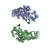
 Mass: 18.015 Da / Num. of mol.: 162 / Source method: isolated from a natural source / Formula: H2O
 Sample preparation
 / Beamline: ID14-2 / Wavelength: 0.933
  Processing