 Movie
Movie Controller


+ Open data

- Basic information
| Entry | Database: PDB / ID: 2zwz | ||||||
|---|---|---|---|---|---|---|---|
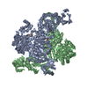
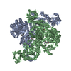
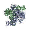
| Title | alpha-L-fucosidase complexed with inhibitor, Core1 | ||||||
 Components Components | Alpha-L-fucosidase, putative | ||||||
 Keywords Keywords | HYDROLASE / Tim Barrel | ||||||
| Function / homology |  Function and homology information Function and homology informationalpha-L-fucosidase / alpha-L-fucosidase activity / fucose metabolic process / glycoside catabolic process / lysosome Similarity search - Function | ||||||
| Biological species |   Thermotoga maritima (bacteria) Thermotoga maritima (bacteria) | ||||||
| Method |  X-RAY DIFFRACTION / SYNCHROTRON / MOLECULAR REPLACEMENT / Resolution: 2.36 Å X-RAY DIFFRACTION / SYNCHROTRON / MOLECULAR REPLACEMENT / Resolution: 2.36 Å | ||||||
 Authors Authors | Wu, H.-J. / Ko, T.-P. / Ho, C.-W. / Lin, C.-H. / Wang, A.H.-J. | ||||||
 Citation Citation | Journal: Angew.Chem.Int.Ed.Engl. / Year: 2010 Title: Structural basis of alpha-fucosidase inhibition by iminocyclitols with K(i) values in the micro- to picomolar range. Authors: Wu, H.J. / Ho, C.W. / Ko, T.P. / Popat, S.D. / Lin, C.H. / Wang, A.H. | ||||||
| History |
|
- Structure visualization
| Structure viewer | Molecule: MolmilJmol/JSmol |
|---|
- Downloads & links
-Download
| PDBx/mmCIF format | 2zwz.cif.gz | 199.1 KB | Display | PDBx/mmCIF format |
|---|---|---|---|---|
| PDB format | pdb2zwz.ent.gz | 159.1 KB | Display | PDB format |
| PDBx/mmJSON format | 2zwz.json.gz | Tree view | PDBx/mmJSON format | |
| Others |  Other downloads Other downloads |
-Validation report
| Arichive directory | https://data.pdbj.org/pub/pdb/validation_reports/zw/2zwzftp://data.pdbj.org/pub/pdb/validation_reports/zw/2zwz | HTTPS FTP |
|---|
-Related structure data
| Related structure data |  2zx5C  2zx6C  2zx7C  2zx8C  2zx9C  2zxaC  2zxbC  2zxdC  1hl8S S: Starting model for refinement C: citing same article ( |
|---|---|
| Similar structure data |


-Links
 PDBj
PDBj- Assembly
| Deposited unit | 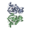
| ||||||||||||||||||
|---|---|---|---|---|---|---|---|---|---|---|---|---|---|---|---|---|---|---|---|
| 1 | 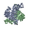
| ||||||||||||||||||
| Unit cell |
| ||||||||||||||||||
| Components on special symmetry positions |
|
-Components
| #1: Protein | Mass: 53100.918 Da / Num. of mol.: 2 Source method: isolated from a genetically manipulated source Source: (gene. exp.) Thermotoga maritima (bacteria) / Strain: MSB8 / Plasmid: pET21b / Production host: #2: Chemical |   Mass: 176.214 Da / Num. of mol.: 2 / Source method: obtained synthetically / Formula: C7H16N2O3 Mass: 176.214 Da / Num. of mol.: 2 / Source method: obtained synthetically / Formula: C7H16N2O3#3: Water | ChemComp-HOH / |  Mass: 18.015 Da / Num. of mol.: 507 / Source method: isolated from a natural source / Formula: H2O Mass: 18.015 Da / Num. of mol.: 507 / Source method: isolated from a natural source / Formula: H2OHas protein modification | Y | |
|---|
-Experimental details
-Experiment
| Experiment | Method: X-RAY DIFFRACTION / Number of used crystals: 1 |
|---|
- Sample preparation
| Crystal | Density Matthews: 2.51 Å3/Da / Density % sol: 50.9 % |
|---|---|
| Crystal grow | Temperature: 298 K / Method: vapor diffusion, sitting drop / pH: 7.4 Details: 14% PEG 5000 MME, 0.1M HEPES, pH 7.4, 4% Jeffamine M-600, VAPOR DIFFUSION, SITTING DROP, temperature 298K |
-Data collection
| Diffraction | Mean temperature: 100 K |
|---|---|
| Diffraction source | Source: SYNCHROTRON / Site: NSRRC  / Beamline: BL13C1 / Wavelength: 0.97315 Å / Beamline: BL13C1 / Wavelength: 0.97315 Å |
| Detector | Type: ADSC QUANTUM 210 / Detector: CCD / Date: Oct 9, 2007 / Details: Vertically focusing mirror |
| Radiation | Monochromator: horizontally focusing single crystal Si(111) bent monochromator Protocol: SINGLE WAVELENGTH / Monochromatic (M) / Laue (L): M / Scattering type: x-ray |
| Radiation wavelength | Wavelength: 0.97315 Å / Relative weight: 1 |
| Reflection | Resolution: 2.36→30 Å / Num. all: 43512 / Num. obs: 43077 / % possible obs: 99 % / Observed criterion σ(F): 0 / Observed criterion σ(I): 1 / Redundancy: 4.8 % / Rmerge(I) obs: 0.07 / Net I/σ(I): 21.4 |
| Reflection shell | Resolution: 2.36→2.44 Å / Redundancy: 4.8 % / Rmerge(I) obs: 0.497 / Mean I/σ(I) obs: 2.9 / Num. unique all: 4281 / % possible all: 99.7 |
- Processing
| Software |
| |||||||||||||||||||||||||
|---|---|---|---|---|---|---|---|---|---|---|---|---|---|---|---|---|---|---|---|---|---|---|---|---|---|---|
| Refinement | Method to determine structure: MOLECULAR REPLACEMENT Starting model: PDB enrty 1HL8 Resolution: 2.36→30 Å / Cross valid method: THROUGHOUT / σ(F): 0 / σ(I): 0 / Stereochemistry target values: Engh & Huber
| |||||||||||||||||||||||||
| Solvent computation | Bsol: 61.256 Å2 | |||||||||||||||||||||||||
| Displacement parameters | Biso max: 102.1 Å2 / Biso mean: 35.125 Å2 / Biso min: 12.88 Å2
| |||||||||||||||||||||||||
| Refine analyze |
| |||||||||||||||||||||||||
| Refinement step | Cycle: LAST / Resolution: 2.36→30 Å
| |||||||||||||||||||||||||
| Refine LS restraints |
| |||||||||||||||||||||||||
| LS refinement shell | Resolution: 2.36→2.44 Å / Rfactor Rfree error: 0.0336
|
