 Movie
Movie Controller

[English] 日本語
 Yorodumi
- PDB-1hl9: CRYSTAL STRUCTURE OF THERMOTOGA MARITIMA ALPHA-FUCOSIDASE IN COMP... -
+ Open data

- Basic information
| Entry | Database: PDB / ID: 1hl9 | ||||||
|---|---|---|---|---|---|---|---|
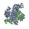
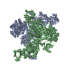
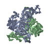
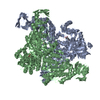
| Title | CRYSTAL STRUCTURE OF THERMOTOGA MARITIMA ALPHA-FUCOSIDASE IN COMPLEX WITH A MECHANISM BASED INHIBITOR | ||||||
 Components Components | PUTATIVE ALPHA-L-FUCOSIDASE | ||||||
 Keywords Keywords | HYDROLASE / GLYCOSIDE HYDROLASE / ALPHA-L-FUCOSIDASE / GLYCOSYL-ENZYME INTERMEDIATE / THERMOSTABLE | ||||||
| Function / homology |  Function and homology information Function and homology informationalpha-L-fucosidase / alpha-L-fucosidase activity / fucose metabolic process / glycoside catabolic process / lysosome Similarity search - Function | ||||||
| Biological species |   THERMOTOGA MARITIMA (bacteria) THERMOTOGA MARITIMA (bacteria) | ||||||
| Method |  X-RAY DIFFRACTION / SYNCHROTRON / MOLECULAR REPLACEMENT / Resolution: 2.25 Å X-RAY DIFFRACTION / SYNCHROTRON / MOLECULAR REPLACEMENT / Resolution: 2.25 Å | ||||||
 Authors Authors | Sulzenbacher, G. / Bignon, C. / Bourne, Y. / Henrissat, B. | ||||||
 Citation Citation | Journal: J.Biol.Chem. / Year: 2004 Title: Crystal Structure of Thermotoga Maritima {Alpha}-L-Fucosidase: Insights Into the Catalytic Mechanism and the Molecular Basis for Fucosidosis Authors: Sulzenbacher, G. / Bignon, C. / Nishimura, T. / Tarling, C.A. / Withers, S.G. / Henrissat, B. / Bourne, Y. | ||||||
| History |
| ||||||
| Remark 650 | HELIX DETERMINATION METHOD: AUTHOR PROVIDED. | ||||||
| Remark 700 | SHEET THE SHEET STRUCTURE OF THIS MOLECULE IS BIFURCATED. IN ORDER TO REPRESENT THIS FEATURE IN ... SHEET THE SHEET STRUCTURE OF THIS MOLECULE IS BIFURCATED. IN ORDER TO REPRESENT THIS FEATURE IN THE SHEET RECORDS BELOW, TWO SHEETS ARE DEFINED. |
- Structure visualization
| Structure viewer | Molecule: MolmilJmol/JSmol |
|---|
- Downloads & links
-Download
| PDBx/mmCIF format | 1hl9.cif.gz | 189.3 KB | Display | PDBx/mmCIF format |
|---|---|---|---|---|
| PDB format | pdb1hl9.ent.gz | 151.3 KB | Display | PDB format |
| PDBx/mmJSON format | 1hl9.json.gz | Tree view | PDBx/mmJSON format | |
| Others |  Other downloads Other downloads |
-Validation report
| Arichive directory | https://data.pdbj.org/pub/pdb/validation_reports/hl/1hl9ftp://data.pdbj.org/pub/pdb/validation_reports/hl/1hl9 | HTTPS FTP |
|---|
-Related structure data
| Related structure data |  1hl8SC  1oduC S: Starting model for refinement C: citing same article ( |
|---|---|
| Similar structure data |

-Links
 PDBj
PDBj- Assembly
| Deposited unit | 
| |||||||||
|---|---|---|---|---|---|---|---|---|---|---|
| 1 | 
| |||||||||
| Unit cell |
| |||||||||
| Components on special symmetry positions |
| |||||||||
| Noncrystallographic symmetry (NCS) | NCS oper: (Code: given Matrix: (-0.67846, -0.73296, -0.04956), Vector: |
-Components
| #1: Protein | Mass: 52315.051 Da / Num. of mol.: 2 Source method: isolated from a genetically manipulated source Details: ORF TM0306 / Source: (gene. exp.) THERMOTOGA MARITIMA (bacteria) / Strain: MSB8 / Production host: #2: Sugar |   Type: L-saccharide, beta linking / Mass: 166.148 Da / Num. of mol.: 2 / Source method: obtained synthetically / Formula: C6H11FO4 Type: L-saccharide, beta linking / Mass: 166.148 Da / Num. of mol.: 2 / Source method: obtained synthetically / Formula: C6H11FO4#3: Water | ChemComp-HOH / |  Mass: 18.015 Da / Num. of mol.: 209 / Source method: isolated from a natural source / Formula: H2O Mass: 18.015 Da / Num. of mol.: 209 / Source method: isolated from a natural source / Formula: H2OCompound details | ALPHA-L-FUCOSIDASE BELONGS TO GLYCOSYL HYDROLASASE FAMILY 29. THE ENZYME PERFORMS CATALYSIS WITH ...ALPHA-L-FUCOSIDASE | |
|---|
-Experimental details
-Experiment
| Experiment | Method: X-RAY DIFFRACTION / Number of used crystals: 1 |
|---|
- Sample preparation
| Crystal | Density Matthews: 2.34 Å3/Da / Density % sol: 47 % |
|---|---|
| Crystal grow | pH: 8 Details: 18% PEG600, 5% JEFFAMINE M600, 100 MM TRIS-HCL PH 8.0, PROTEIN CONCENTRATION 5 MG/ML THE INHIBITOR WAS INTRODUCED BY SHORT SOAKING OF A NATIVE |
-Data collection
| Diffraction | Mean temperature: 100 K |
|---|---|
| Diffraction source | Source: SYNCHROTRON / Site: ESRF  / Beamline: ID14-1 / Wavelength: 0.934 / Beamline: ID14-1 / Wavelength: 0.934 |
| Detector | Type: ADSC CCD / Detector: CCD / Date: Mar 11, 2002 |
| Radiation | Protocol: SINGLE WAVELENGTH / Monochromatic (M) / Laue (L): M / Scattering type: x-ray |
| Radiation wavelength | Wavelength: 0.934 Å / Relative weight: 1 |
| Reflection | Resolution: 2.25→55.9 Å / Num. obs: 37238 / % possible obs: 89.4 % / Redundancy: 4.2 % / Rmerge(I) obs: 0.065 / Net I/σ(I): 7.1 |
| Reflection shell | Resolution: 2.25→2.31 Å / Redundancy: 4 % / Rmerge(I) obs: 0.397 / Mean I/σ(I) obs: 1.8 / % possible all: 89.4 |
- Processing
| Software |
| ||||||||||||||||||||||||||||||||||||||||||||||||||||||||||||||||||||||||||||||||||||||||||||||||||||||||||||||||||||||||||||||||||||||||||||||||||||||||||||||||||||||||||||||||||||||
|---|---|---|---|---|---|---|---|---|---|---|---|---|---|---|---|---|---|---|---|---|---|---|---|---|---|---|---|---|---|---|---|---|---|---|---|---|---|---|---|---|---|---|---|---|---|---|---|---|---|---|---|---|---|---|---|---|---|---|---|---|---|---|---|---|---|---|---|---|---|---|---|---|---|---|---|---|---|---|---|---|---|---|---|---|---|---|---|---|---|---|---|---|---|---|---|---|---|---|---|---|---|---|---|---|---|---|---|---|---|---|---|---|---|---|---|---|---|---|---|---|---|---|---|---|---|---|---|---|---|---|---|---|---|---|---|---|---|---|---|---|---|---|---|---|---|---|---|---|---|---|---|---|---|---|---|---|---|---|---|---|---|---|---|---|---|---|---|---|---|---|---|---|---|---|---|---|---|---|---|---|---|---|---|
| Refinement | Method to determine structure: MOLECULAR REPLACEMENT Starting model: PDB ENTRY 1HL8 Resolution: 2.25→20 Å / Cor.coef. Fo:Fc: 0.957 / Cor.coef. Fo:Fc free: 0.936 / SU B: 6.362 / SU ML: 0.159 / TLS residual ADP flag: LIKELY RESIDUAL / Cross valid method: THROUGHOUT / ESU R: 0.411 / ESU R Free: 0.242 / Stereochemistry target values: MAXIMUM LIKELIHOOD Details: HYDROGENS HAVE BEEN ADDED IN THE RIDING POSITIONS. TWO OVERLAPPING SPECIES ARE PRESENT IN THE ACTIVE SITE AND HAVE BEEN REFINED WITH PARTIAL OCCUPANCIES.
| ||||||||||||||||||||||||||||||||||||||||||||||||||||||||||||||||||||||||||||||||||||||||||||||||||||||||||||||||||||||||||||||||||||||||||||||||||||||||||||||||||||||||||||||||||||||
| Solvent computation | Ion probe radii: 0.8 Å / Shrinkage radii: 0.8 Å / VDW probe radii: 1.4 Å / Solvent model: BABINET MODEL WITH MASK | ||||||||||||||||||||||||||||||||||||||||||||||||||||||||||||||||||||||||||||||||||||||||||||||||||||||||||||||||||||||||||||||||||||||||||||||||||||||||||||||||||||||||||||||||||||||
| Displacement parameters | Biso mean: 24.26 Å2
| ||||||||||||||||||||||||||||||||||||||||||||||||||||||||||||||||||||||||||||||||||||||||||||||||||||||||||||||||||||||||||||||||||||||||||||||||||||||||||||||||||||||||||||||||||||||
| Refinement step | Cycle: LAST / Resolution: 2.25→20 Å
| ||||||||||||||||||||||||||||||||||||||||||||||||||||||||||||||||||||||||||||||||||||||||||||||||||||||||||||||||||||||||||||||||||||||||||||||||||||||||||||||||||||||||||||||||||||||
| Refine LS restraints |
|