 Movie
Movie Controller

[English] 日本語
 Yorodumi
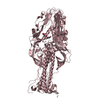
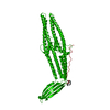
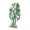
- PDB-2vde: Crystal Structure of the Open State of TolC Outer Membrane Compon... -
+ Open data

- Basic information
| Entry | Database: PDB / ID: 2vde | ||||||
|---|---|---|---|---|---|---|---|
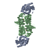
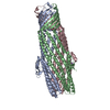
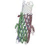
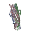
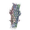
| Title | Crystal Structure of the Open State of TolC Outer Membrane Component of Mutlidrug Efflux Pumps | ||||||
 Components Components | OUTER MEMBRANE PROTEIN TOLC | ||||||
 Keywords Keywords | TRANSPORT PROTEIN / OUTER MEMBRANE / ALPHA HELICAL BARREL / MULTIDRUG EFFLUX PUMP / INTEGRAL MEMBRANE PROTEIN / MEMBRANE / TRANSPORT / BETA BARREL / TRANSMEMBRANE | ||||||
| Function / homology |  Function and homology information Function and homology informationMacAB-TolC complex / enterobactin transport / enterobactin transmembrane transporter activity / xenobiotic detoxification by transmembrane export across the cell outer membrane / periplasmic side of plasma membrane / efflux pump complex / Iron assimilation using enterobactin / bile acid transmembrane transporter activity / xenobiotic detoxification by transmembrane export across the plasma membrane / Antimicrobial resistance ...MacAB-TolC complex / enterobactin transport / enterobactin transmembrane transporter activity / xenobiotic detoxification by transmembrane export across the cell outer membrane / periplasmic side of plasma membrane / efflux pump complex / Iron assimilation using enterobactin / bile acid transmembrane transporter activity / xenobiotic detoxification by transmembrane export across the plasma membrane / Antimicrobial resistance / bile acid and bile salt transport / porin activity / Secretion of toxins / efflux transmembrane transporter activity / monoatomic ion channel activity / cell outer membrane / response to toxic substance / monoatomic ion transmembrane transport / response to xenobiotic stimulus / response to antibiotic / membrane / identical protein binding Similarity search - Function | ||||||
| Biological species |  | ||||||
| Method |  X-RAY DIFFRACTION / SYNCHROTRON / MOLECULAR REPLACEMENT / Resolution: 3.2 Å X-RAY DIFFRACTION / SYNCHROTRON / MOLECULAR REPLACEMENT / Resolution: 3.2 Å | ||||||
 Authors Authors | Bavro, V.N. / Pietras, Z. / Furnham, N. / Perez-Cano, L. / Fernandez-Recio, J. / Pei, X.Y. / Truer, R. / Misra, R. / Luisi, B. | ||||||
 Citation Citation | Journal: Mol.Cell / Year: 2008 Title: Assembly and Channel Opening in a Bacterial Drug Efflux Machine. Authors: Bavro, V.N. / Pietras, Z. / Furnham, N. / Perez-Cano, L. / Fernandez-Recio, J. / Pei, X.Y. / Truer, R. / Misra, R. / Luisi, B. | ||||||
| History |
| ||||||
| Remark 650 | HELIX DETERMINATION METHOD: AUTHOR PROVIDED. | ||||||
| Remark 700 | SHEET DETERMINATION METHOD: AUTHOR PROVIDED. |
- Structure visualization
| Structure viewer | Molecule: MolmilJmol/JSmol |
|---|
- Downloads & links
-Download
| PDBx/mmCIF format | 2vde.cif.gz | 249.3 KB | Display | PDBx/mmCIF format |
|---|---|---|---|---|
| PDB format | pdb2vde.ent.gz | 202 KB | Display | PDB format |
| PDBx/mmJSON format | 2vde.json.gz | Tree view | PDBx/mmJSON format | |
| Others |  Other downloads Other downloads |
-Validation report
| Arichive directory | https://data.pdbj.org/pub/pdb/validation_reports/vd/2vdeftp://data.pdbj.org/pub/pdb/validation_reports/vd/2vde | HTTPS FTP |
|---|
-Related structure data
| Related structure data | 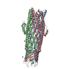 2vddC 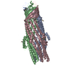 1ek9S S: Starting model for refinement C: citing same article ( |
|---|---|
| Similar structure data |


-Links
 PDBj
PDBj
- Assembly
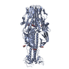
| Deposited unit | 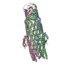
| ||||||||
|---|---|---|---|---|---|---|---|---|---|
| 1 |
| ||||||||
| Unit cell |
|
-Components
| #1: Protein | Mass: 50623.000 Da / Num. of mol.: 3 / Fragment: RESIDUES 1-450 / Mutation: YES Source method: isolated from a genetically manipulated source Source: (gene. exp.) #2: Chemical | ChemComp-CL /   Mass: 35.453 Da / Num. of mol.: 4 / Source method: obtained synthetically / Formula: Cl Mass: 35.453 Da / Num. of mol.: 4 / Source method: obtained synthetically / Formula: ClCompound details | ENGINEERED RESIDUE IN CHAIN A, VAL 191 TO LEU ENGINEERED RESIDUE IN CHAIN A, TYR 384 TO PHE ...ENGINEERED | Nonpolymer details | CHLORIDE ION (CL): CHLORIDE DEDUCED FROM COORDINATI | |
|---|
-Experimental details
-Experiment
| Experiment | Method: X-RAY DIFFRACTION / Number of used crystals: 6 |
|---|
- Sample preparation
| Crystal | Density Matthews: 3.83 Å3/Da / Density % sol: 67.91 % / Description: NONE |
|---|---|
| Crystal grow | pH: 10.5 Details: PROTEIN WAS CRYSTALLIZED FROM 0.1M CAPS PH10.5, 30% PEG400 |
-Data collection
| Diffraction | Mean temperature: 100 K |
|---|---|
| Diffraction source | Source: SYNCHROTRON / Site: ESRF  / Beamline: ID23-2 / Wavelength: 0.8726 / Beamline: ID23-2 / Wavelength: 0.8726 |
| Detector | Type: MARRESEARCH / Detector: CCD / Date: Feb 10, 2007 / Details: SLITS |
| Radiation | Monochromator: HORIZONTALLY SIDE DIFFRACTING SILICON 111 CRYSTAL Protocol: SINGLE WAVELENGTH / Monochromatic (M) / Laue (L): M / Scattering type: x-ray |
| Radiation wavelength | Wavelength: 0.8726 Å / Relative weight: 1 |
| Reflection | Resolution: 3.2→22.1 Å / Num. obs: 36034 / % possible obs: 97.7 % / Observed criterion σ(I): 2.4 / Redundancy: 5 % / Rmerge(I) obs: 0.15 / Net I/σ(I): 9.53 |
| Reflection shell | Resolution: 3.2→3.3 Å / Redundancy: 5 % / Rmerge(I) obs: 0.42 / Mean I/σ(I) obs: 2.45 / % possible all: 98.3 |
- Processing
| Software |
| ||||||||||||||||||||||||||||||||||||||||||||||||||||||||||||||||||||||||||||||||||||||||||||||||||||||||||||||||||||||||||||||||||||||||||||||||||||||||||||||||||||||||||||||||||||||
|---|---|---|---|---|---|---|---|---|---|---|---|---|---|---|---|---|---|---|---|---|---|---|---|---|---|---|---|---|---|---|---|---|---|---|---|---|---|---|---|---|---|---|---|---|---|---|---|---|---|---|---|---|---|---|---|---|---|---|---|---|---|---|---|---|---|---|---|---|---|---|---|---|---|---|---|---|---|---|---|---|---|---|---|---|---|---|---|---|---|---|---|---|---|---|---|---|---|---|---|---|---|---|---|---|---|---|---|---|---|---|---|---|---|---|---|---|---|---|---|---|---|---|---|---|---|---|---|---|---|---|---|---|---|---|---|---|---|---|---|---|---|---|---|---|---|---|---|---|---|---|---|---|---|---|---|---|---|---|---|---|---|---|---|---|---|---|---|---|---|---|---|---|---|---|---|---|---|---|---|---|---|---|---|
| Refinement | Method to determine structure: MOLECULAR REPLACEMENT Starting model: PDB ENTRY 1EK9 Resolution: 3.2→22.08 Å / Cor.coef. Fo:Fc: 0.885 / Cor.coef. Fo:Fc free: 0.839 / Cross valid method: THROUGHOUT / ESU R Free: 0.541 / Stereochemistry target values: MAXIMUM LIKELIHOOD Details: HYDROGENS HAVE BEEN ADDED IN THE RIDING POSITIONS. DISORDERED REGIONS WERE MODELED IN FULL WITH OCCUPANCY ZERO.
| ||||||||||||||||||||||||||||||||||||||||||||||||||||||||||||||||||||||||||||||||||||||||||||||||||||||||||||||||||||||||||||||||||||||||||||||||||||||||||||||||||||||||||||||||||||||
| Solvent computation | Ion probe radii: 0.8 Å / Shrinkage radii: 0.8 Å / VDW probe radii: 1.2 Å / Solvent model: MASK | ||||||||||||||||||||||||||||||||||||||||||||||||||||||||||||||||||||||||||||||||||||||||||||||||||||||||||||||||||||||||||||||||||||||||||||||||||||||||||||||||||||||||||||||||||||||
| Displacement parameters | Biso mean: 67.82 Å2
| ||||||||||||||||||||||||||||||||||||||||||||||||||||||||||||||||||||||||||||||||||||||||||||||||||||||||||||||||||||||||||||||||||||||||||||||||||||||||||||||||||||||||||||||||||||||
| Refinement step | Cycle: LAST / Resolution: 3.2→22.08 Å
| ||||||||||||||||||||||||||||||||||||||||||||||||||||||||||||||||||||||||||||||||||||||||||||||||||||||||||||||||||||||||||||||||||||||||||||||||||||||||||||||||||||||||||||||||||||||
| Refine LS restraints |
|