[English] 日本語
Yorodumi
- PDB-4k7r: Crystal structures of CusC review conformational changes accompan... -

+
Open data


ID or keywords:

Loading...

-
Basic information

Entry
Database: PDB / ID: 4k7r
Title
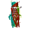
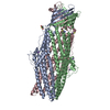
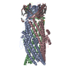
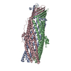
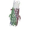
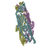
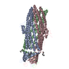
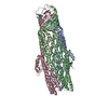
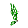
Crystal structures of CusC review conformational changes accompanying folding and transmembrane channel formation
ComponentsCation efflux system protein CusC
KeywordsMEMBRANE PROTEIN / beta barrel
Function / homology
Function and homology information


protein palmitoylation / copper ion transmembrane transport / response to silver ion / silver ion transmembrane transport / diacylglycerol binding / copper ion transmembrane transporter activity / copper ion export / detoxification of copper ion / response to copper ion / porin activity ...protein palmitoylation / copper ion transmembrane transport / response to silver ion / silver ion transmembrane transport / diacylglycerol binding / copper ion transmembrane transporter activity / copper ion export / detoxification of copper ion / response to copper ion / porin activity / pore complex / protein homotrimerization / efflux transmembrane transporter activity / transmembrane transporter activity / intracellular copper ion homeostasis / cell outer membrane / response to toxic substance / transmembrane transport / copper ion binding / identical protein binding / membrane
Similarity search - Function
: / Outer membrane efflux proteins (OEP) / Outer membrane efflux proteins (OEP) / Outer membrane efflux proteins (OEP) / Outer membrane efflux proteins (OEP) / RND efflux system, outer membrane lipoprotein, NodT / Outer membrane efflux protein / Outer membrane efflux protein / Single Sheet / Prokaryotic membrane lipoprotein lipid attachment site profile. ...: / Outer membrane efflux proteins (OEP) / Outer membrane efflux proteins (OEP) / Outer membrane efflux proteins (OEP) / Outer membrane efflux proteins (OEP) / RND efflux system, outer membrane lipoprotein, NodT / Outer membrane efflux protein / Outer membrane efflux protein / Single Sheet / Prokaryotic membrane lipoprotein lipid attachment site profile. / Up-down Bundle / Mainly Beta / Mainly Alpha
Similarity search - Domain/homology
(2S)-1-(pentanoyloxy)propan-2-yl hexanoate / Cation efflux system protein CusC
Similarity search - Component
Biological speciesEscherichia coli (E. coli)
MethodX-RAY DIFFRACTION / SYNCHROTRON / MOLECULAR REPLACEMENT / Resolution: 2.094 Å
AuthorsSu, C.-C. / Lei, H.-T. / Bolla, J.R. / Yu, E.W.
CitationJournal: J.Mol.Biol. / Year: 2014
Title: Crystal Structures of CusC Review Conformational Changes Accompanying Folding and Transmembrane Channel Formation.
Authors: Lei, H.T. / Bolla, J.R. / Bishop, N.R. / Su, C.C. / Yu, E.W.
History
DepositionApr 17, 2013Deposition site: RCSB / Processing site: RCSB
Revision 1.0Oct 16, 2013Provider: repository / Type: Initial release
Revision 1.1Feb 5, 2014Group: Database references
Revision 1.2Feb 28, 2024Group: Data collection / Database references / Derived calculations
Category: chem_comp_atom / chem_comp_bond ...chem_comp_atom / chem_comp_bond / database_2 / struct_ref_seq_dif / struct_site
Item: _database_2.pdbx_DOI / _database_2.pdbx_database_accession ..._database_2.pdbx_DOI / _database_2.pdbx_database_accession / _struct_ref_seq_dif.details / _struct_site.pdbx_auth_asym_id / _struct_site.pdbx_auth_comp_id / _struct_site.pdbx_auth_seq_id

-
Structure visualization

Structure viewerMolecule:
MolmilJmol/JSmol

Downloads & links

-
Assembly

Deposited unit
A: Cation efflux system protein CusC
hetero molecules


Theoretical massNumber of molelcules
Total (without water)49,6502
Polymers49,3921
Non-polymers2581
Water5,224290
1
A: Cation efflux system protein CusC
hetero molecules

A: Cation efflux system protein CusC
hetero molecules

A: Cation efflux system protein CusC
hetero molecules


Theoretical massNumber of molelcules
Total (without water)148,9516
Polymers148,1763
Non-polymers7753
Water543
TypeNameSymmetry operationNumber
identity operation1_555x,y,z1
crystal symmetry operation2_565-y,x-y+1,z1
crystal symmetry operation3_455-x+y-1,-x,z1
Buried area16700 Å2
ΔGint-96 kcal/mol
Surface area58870 Å2
MethodPISA
Unit cell
Length a, b, c (Å)89.026, 89.026, 474.103
Angle α, β, γ (deg.)90.000, 90.000, 120.000
Int Tables number155
Space group name H-MH32

-
Components

#1: Protein Cation efflux system protein CusC


Mass: 49391.977 Da / Num. of mol.: 1
Source method: isolated from a genetically manipulated source
Source: (gene. exp.) Escherichia coli (E. coli) / Strain: K-12 / Gene: b0572, cusC, ibeB, JW0561, ylcB / Plasmid: pBAD22 / Production host: Escherichia coli (E. coli) / References: UniProt: P77211
#2: Chemical ChemComp-3PK / (2S)-1-(pentanoyloxy)propan-2-yl hexanoate


Mass: 258.354 Da / Num. of mol.: 1 / Source method: obtained synthetically / Formula: C14H26O4
#3: Water ChemComp-HOH / water


Mass: 18.015 Da / Num. of mol.: 290 / Source method: isolated from a natural source / Formula: H2O
Nonpolymer detailsTHE AUTHORS HAVE IDENTIFIED THE LIGAND HAS UNKNOWN. THE CLOSER MATCH IS THE LIGAND (2S)-1- ...THE AUTHORS HAVE IDENTIFIED THE LIGAND HAS UNKNOWN. THE CLOSER MATCH IS THE LIGAND (2S)-1-(PENTANOYLOXY)PROPAN-2-YL HEXANOATE

-
Experimental details

-
Experiment

ExperimentMethod: X-RAY DIFFRACTION / Number of used crystals: 1

-
Sample preparation

CrystalDensity Matthews: 3.66 Å3/Da / Density % sol: 66.4 %
Crystal growTemperature: 298 K / Method: vapor diffusion / pH: 7.5
Details: 7% PEG3350, 0.2M NH4SO4, 0.1M HEPES, pH 7.5, VAPOR DIFFUSION, temperature 298K

-
Data collection

DiffractionMean temperature: 100 K
Diffraction sourceSource: SYNCHROTRON / Site: APS / Beamline: 24-ID-E / Wavelength: 0.98 Å
DetectorType: ACSD / Detector: CCD / Date: Feb 3, 2012
RadiationMonochromator: Si(III) / Protocol: SINGLE WAVELENGTH / Monochromatic (M) / Laue (L): M / Scattering type: x-ray
Radiation wavelengthWavelength: 0.98 Å / Relative weight: 1
ReflectionResolution: 2.09→50 Å / Num. all: 80183 / Num. obs: 80183 / % possible obs: 97.3 % / Observed criterion σ(F): 2 / Observed criterion σ(I): 2 / Redundancy: 2.4 % / Rmerge(I) obs: 0.087 / Χ2: 1.438 / Net I/σ(I): 9
Reflection shell
Resolution (Å)Redundancy (%)Rmerge(I) obsNum. unique allΧ2Diffraction-ID% possible all
2.09-2.172.40.40489460.827197.2
2.17-2.272.40.30389230.913197.6
2.27-2.392.40.22889590.941197.9
2.39-2.542.40.18489750.986198.1
2.54-2.742.40.13689471.062198.1
2.74-3.012.40.08990151.261198.1
3.01-3.452.40.08288522.031197.1
3.45-4.352.40.05987422.951195.3
4.35-502.50.0488242.029196.2

-
Processing

Software
NameVersionClassificationNB
SCALEPACKdata scaling
PHENIX1.7.3_928refinement
PDB_EXTRACT3.11data extraction
ADSCQuantumdata collection
DENZOdata reduction
PHASERphasing
RefinementMethod to determine structure: MOLECULAR REPLACEMENT / Resolution: 2.094→35.711 Å / Occupancy max: 1 / Occupancy min: 1 / FOM work R set: 0.8347 / SU ML: 0.18 / σ(F): 1.33 / Phase error: 23.37 / Stereochemistry target values: ML
RfactorNum. reflection% reflectionSelection details
Rfree0.25 2189 5.15 %RANDOM
Rwork0.2139 ---
obs0.2157 42478 97.79 %-
all-42478 --
Solvent computationShrinkage radii: 0.86 Å / VDW probe radii: 1.1 Å / Solvent model: FLAT BULK SOLVENT MODEL / Bsol: 58.183 Å2 / ksol: 0.4 e/Å3
Displacement parametersBiso max: 76 Å2 / Biso mean: 31.8148 Å2 / Biso min: 17.07 Å2
Baniso -1Baniso -2Baniso -3
1-6.0876 Å2-0 Å2-0 Å2
2--6.0876 Å2-0 Å2
3----12.1751 Å2
Refinement stepCycle: LAST / Resolution: 2.094→35.711 Å
ProteinNucleic acidLigandSolventTotal
Num. atoms3343 0 18 290 3651
Refine LS restraints
Refine-IDTypeDev idealNumber
X-RAY DIFFRACTIONf_bond_d0.0073410
X-RAY DIFFRACTIONf_angle_d0.9514614
X-RAY DIFFRACTIONf_chiral_restr0.06519
X-RAY DIFFRACTIONf_plane_restr0.004612
X-RAY DIFFRACTIONf_dihedral_angle_d15.2271267
LS refinement shell

Refine-ID: X-RAY DIFFRACTION / Total num. of bins used: 16

Resolution (Å)Rfactor RfreeNum. reflection RfreeRfactor RworkNum. reflection RworkNum. reflection all% reflection obs (%)
2.094-2.13950.29371350.24742473260898
2.1395-2.18930.27441560.23124912647100
2.1893-2.2440.27271400.221425472687100
2.244-2.30470.26391390.208325182657100
2.3047-2.37250.21591340.202725382672100
2.3725-2.44910.27191300.20325512681100
2.4491-2.53660.21541370.197925472684100
2.5366-2.63810.21891440.19542516266099
2.6381-2.75810.25971500.20672504265499
2.7581-2.90350.26161210.21082532265398
2.9035-3.08530.28811310.21152539267098
3.0853-3.32340.25931420.21312480262297
3.3234-3.65750.25191290.21822462259195
3.6575-4.18610.22981350.18952433256894
4.1861-5.27130.24131350.19912532266796
5.2713-35.71620.2441310.26572626275794

+
About Yorodumi

-
News

-
Feb 9, 2022. New format data for meta-information of EMDB entries

New format data for meta-information of EMDB entries

  • Version 3 of the EMDB header file is now the official format.
  • The previous official version 1.9 will be removed from the archive.

Related info.:EMDB header

External links:wwPDB to switch to version 3 of the EMDB data model

-
Aug 12, 2020. Covid-19 info

Covid-19 info

URL: https://pdbjlvh1.pdbj.org/emnavi/covid19.php

New page: Covid-19 featured information page in EM Navigator.

Related info.:Covid-19 info / Mar 5, 2020. Novel coronavirus structure data

+
Mar 5, 2020. Novel coronavirus structure data

Novel coronavirus structure data

Related info.:Yorodumi Speices / Aug 12, 2020. Covid-19 info

External links:COVID-19 featured content - PDBj / Molecule of the Month (242):Coronavirus Proteases

+
Jan 31, 2019. EMDB accession codes are about to change! (news from PDBe EMDB page)

EMDB accession codes are about to change! (news from PDBe EMDB page)

  • The allocation of 4 digits for EMDB accession codes will soon come to an end. Whilst these codes will remain in use, new EMDB accession codes will include an additional digit and will expand incrementally as the available range of codes is exhausted. The current 4-digit format prefixed with “EMD-” (i.e. EMD-XXXX) will advance to a 5-digit format (i.e. EMD-XXXXX), and so on. It is currently estimated that the 4-digit codes will be depleted around Spring 2019, at which point the 5-digit format will come into force.
  • The EM Navigator/Yorodumi systems omit the EMD- prefix.

Related info.:Q: What is EMD? / ID/Accession-code notation in Yorodumi/EM Navigator

External links:EMDB Accession Codes are Changing Soon! / Contact to PDBj

+
Jul 12, 2017. Major update of PDB

Major update of PDB

  • wwPDB released updated PDB data conforming to the new PDBx/mmCIF dictionary.
  • This is a major update changing the version number from 4 to 5, and with Remediation, in which all the entries are updated.
  • In this update, many items about electron microscopy experimental information are reorganized (e.g. em_software).
  • Now, EM Navigator and Yorodumi are based on the updated data.

External links:wwPDB Remediation / Enriched Model Files Conforming to OneDep Data Standards Now Available in the PDB FTP Archive

-
Yorodumi

Thousand views of thousand structures

  • Yorodumi is a browser for structure data from EMDB, PDB, SASBDB, etc.
  • This page is also the successor to EM Navigator detail page, and also detail information page/front-end page for Omokage search.
  • The word "yorodu" (or yorozu) is an old Japanese word meaning "ten thousand". "mi" (miru) is to see.

Related info.:EMDB / PDB / SASBDB / Comparison of 3 databanks / Yorodumi Search / Aug 31, 2016. New EM Navigator & Yorodumi / Yorodumi Papers / Jmol/JSmol / Function and homology information / Changes in new EM Navigator and Yorodumi

Read more