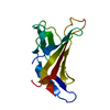
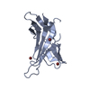
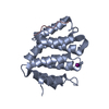
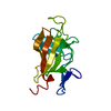
[English] 日本語
Yorodumi
- PDB-2v5o: STRUCTURE OF HUMAN IGF2R DOMAINS 11-14 -

+
Open data


ID or keywords:

Loading...

-
Basic information

Entry
Database: PDB / ID: 2v5o
TitleSTRUCTURE OF HUMAN IGF2R DOMAINS 11-14
ComponentsCATION-INDEPENDENT MANNOSE-6-PHOSPHATE RECEPTOR
KeywordsRECEPTOR / CATION INDEPENDENT MANNOSE 6-PHOSPHATE / MEMBRANE / LYSOSOME / TRANSPORT / BETA BARREL / PHOSPHORYLATION / FIBRONECTIN TYPE II / INSULIN-LIKE GROWTH FACTOR / GLYCOPROTEIN / TRANSMEMBRANE
Function / homology
Function and homology information


Retrograde transport at the Trans-Golgi-Network / retromer complex binding / clathrin coat / response to tetrachloromethane / insulin-like growth factor receptor activity / insulin-like growth factor binding / positive regulation by host of viral process / insulin-like growth factor II binding / trans-Golgi network transport vesicle / retinoic acid binding ...Retrograde transport at the Trans-Golgi-Network / retromer complex binding / clathrin coat / response to tetrachloromethane / insulin-like growth factor receptor activity / insulin-like growth factor binding / positive regulation by host of viral process / insulin-like growth factor II binding / trans-Golgi network transport vesicle / retinoic acid binding / lysosomal transport / Golgi Associated Vesicle Biogenesis / nuclear envelope lumen / mannose binding / endocytic vesicle / G-protein alpha-subunit binding / animal organ regeneration / response to retinoic acid / transport vesicle / receptor-mediated endocytosis / post-embryonic development / secretory granule membrane / trans-Golgi network membrane / liver development / phosphoprotein binding / clathrin-coated endocytic vesicle membrane / trans-Golgi network / late endosome / Cargo recognition for clathrin-mediated endocytosis / signaling receptor activity / Clathrin-mediated endocytosis / spermatogenesis / early endosome / endosome membrane / endosome / positive regulation of apoptotic process / G protein-coupled receptor signaling pathway / Golgi membrane / focal adhesion / Neutrophil degranulation / perinuclear region of cytoplasm / Golgi apparatus / enzyme binding / cell surface / signal transduction / extracellular exosome / membrane / identical protein binding / plasma membrane
Similarity search - Function
Fibronectin, type II, collagen-binding / Cation-independent mannose-6-phosphate receptor repeat / Cation-dependent Mannose-6-phosphate Receptor; Chain A / Mannose-6-phosphate receptor binding domain / Cation-independent mannose-6-phosphate receptor repeat / Cation-independent mannose-6-phosphate receptor repeat / MRH domain / MRH domain profile. / Mannose-6-phosphate receptor binding domain superfamily / Seminal Fluid Protein PDC-109 (Domain B) ...Fibronectin, type II, collagen-binding / Cation-independent mannose-6-phosphate receptor repeat / Cation-dependent Mannose-6-phosphate Receptor; Chain A / Mannose-6-phosphate receptor binding domain / Cation-independent mannose-6-phosphate receptor repeat / Cation-independent mannose-6-phosphate receptor repeat / MRH domain / MRH domain profile. / Mannose-6-phosphate receptor binding domain superfamily / Seminal Fluid Protein PDC-109 (Domain B) / Fibronectin type II domain / Fibronectin type II domain superfamily / Fibronectin type II domain / Fibronectin type-II collagen-binding domain signature. / Fibronectin type-II collagen-binding domain profile. / Fibronectin type 2 domain / Kringle-like fold / Distorted Sandwich / Ribbon / Mainly Beta
Similarity search - Domain/homology
Cation-independent mannose-6-phosphate receptor
Similarity search - Component
Biological speciesHomo sapiens (human)
MethodX-RAY DIFFRACTION / SYNCHROTRON / MOLECULAR REPLACEMENT / Resolution: 2.91 Å
AuthorsBrown, J. / Delaine, C. / Zaccheo, O.J. / Siebold, C. / Gilbert, R.J. / van Boxel, G. / Denley, A. / Wallace, J.C. / Hassan, A.B. / Forbes, B.E. / Jones, E.Y.
CitationJournal: Embo J. / Year: 2008
Title: Structure and Functional Analysis of the Igf-II/Igf2R Interaction
Authors: Brown, J. / Delaine, C. / Zaccheo, O.J. / Siebold, C. / Gilbert, R.J. / Van Boxel, G. / Denley, A. / Wallace, J.C. / Hassan, A.B. / Forbes, B.E. / Jones, E.Y.
History
DepositionJul 6, 2007Deposition site: PDBE / Processing site: PDBE
Revision 1.0Dec 11, 2007Provider: repository / Type: Initial release
Revision 1.1Jul 13, 2011Group: Advisory / Version format compliance
Revision 1.2Feb 28, 2018Group: Source and taxonomy / Category: entity_src_gen
Item: _entity_src_gen.gene_src_common_name / _entity_src_gen.pdbx_gene_src_scientific_name ..._entity_src_gen.gene_src_common_name / _entity_src_gen.pdbx_gene_src_scientific_name / _entity_src_gen.pdbx_host_org_cell_line / _entity_src_gen.pdbx_host_org_scientific_name / _entity_src_gen.pdbx_host_org_strain / _entity_src_gen.pdbx_host_org_variant
Revision 1.3Jul 29, 2020Group: Data collection / Derived calculations ...Data collection / Derived calculations / Other / Structure summary
Category: chem_comp / entity ...chem_comp / entity / pdbx_chem_comp_identifier / pdbx_database_status / pdbx_entity_nonpoly / struct_conn / struct_site / struct_site_gen
Item: _chem_comp.name / _chem_comp.type ..._chem_comp.name / _chem_comp.type / _entity.pdbx_description / _pdbx_database_status.status_code_sf / _pdbx_entity_nonpoly.name / _struct_conn.pdbx_leaving_atom_flag / _struct_conn.pdbx_role
Description: Carbohydrate remediation / Provider: repository / Type: Remediation
Revision 1.4Dec 13, 2023Group: Data collection / Database references ...Data collection / Database references / Refinement description / Structure summary
Category: chem_comp / chem_comp_atom ...chem_comp / chem_comp_atom / chem_comp_bond / database_2 / pdbx_initial_refinement_model
Item: _chem_comp.pdbx_synonyms / _database_2.pdbx_DOI / _database_2.pdbx_database_accession

-
Structure visualization

Structure viewerMolecule:
MolmilJmol/JSmol

Downloads & links

-
Assembly

Deposited unit
A: CATION-INDEPENDENT MANNOSE-6-PHOSPHATE RECEPTOR
hetero molecules


Theoretical massNumber of molelcules
Total (without water)70,0696
Polymers69,3341
Non-polymers7355
Water0
1


  • Idetical with deposited unit
  • defined by author&software
TypeNameSymmetry operationNumber
identity operation1_555x,y,z1
MethodPQS
Unit cell
Length a, b, c (Å)138.554, 69.247, 98.012
Angle α, β, γ (deg.)90.00, 103.25, 90.00
Int Tables number5
Space group name H-MC121

-
Components

#1: Protein CATION-INDEPENDENT MANNOSE-6-PHOSPHATE RECEPTOR / INSULIN-LIKE GROWTH FACTOR II RECEPTOR / CI MAN-6-P RECEPTOR / CI-MPR / M6PR / INSULIN-LIKE GROWTH ...INSULIN-LIKE GROWTH FACTOR II RECEPTOR / CI MAN-6-P RECEPTOR / CI-MPR / M6PR / INSULIN-LIKE GROWTH FACTOR 2 RECEPTOR / IGF-II RECEPTOR / M6P/IGF2 RECEPTOR / M6P/IGF2R / 300 KDA MANNOSE 6-PHOSPHATE RECEPTOR / MPR 300 / MPR300 / CD222 ANTIGEN


Mass: 69334.406 Da / Num. of mol.: 1 / Fragment: DOMAINS 11-14, RESIDUES 1508-2128
Source method: isolated from a genetically manipulated source
Source: (gene. exp.) Homo sapiens (human) / Organ: PLACENTA / Plasmid: PEE14 / Cell line (production host): CHO / Production host: 
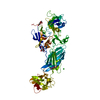
Cricetulus griseus (Chinese hamster) / Variant (production host): LECR 3.2.8.1 / References: UniProt: P11717
#2: Sugar ChemComp-NAG / 2-acetamido-2-deoxy-beta-D-glucopyranose / N-acetyl-beta-D-glucosamine / 2-acetamido-2-deoxy-beta-D-glucose / 2-acetamido-2-deoxy-D-glucose / 2-acetamido-2-deoxy-glucose / N-ACETYL-D-GLUCOSAMINE / N-Acetylglucosamine


Type: D-saccharide, beta linking / Mass: 221.208 Da / Num. of mol.: 3
Source method: isolated from a genetically manipulated source
Formula: C8H15NO6
IdentifierTypeProgram
DGlcpNAcbCONDENSED IUPAC CARBOHYDRATE SYMBOLGMML 1.0
N-acetyl-b-D-glucopyranosamineCOMMON NAMEGMML 1.0
b-D-GlcpNAcIUPAC CARBOHYDRATE SYMBOLPDB-CARE 1.0
GlcNAcSNFG CARBOHYDRATE SYMBOLGMML 1.0
#3: Chemical ChemComp-CL / CHLORIDE ION / Chloride


Mass: 35.453 Da / Num. of mol.: 2 / Source method: obtained synthetically / Formula: Cl
Sequence detailsTHE CONFLICT INDICATED IN THE SEQADV RECORDS BELOW HAS BEEN DESCRIBED IN UNIPROT ENTRY P11717 UNDER ...THE CONFLICT INDICATED IN THE SEQADV RECORDS BELOW HAS BEEN DESCRIBED IN UNIPROT ENTRY P11717 UNDER REFERENCE WITH PUBMEDID: 2957598.

-
Experimental details

-
Experiment

ExperimentMethod: X-RAY DIFFRACTION / Number of used crystals: 1

-
Sample preparation

CrystalDensity Matthews: 3.3 Å3/Da / Density % sol: 63 %
Crystal growpH: 6.4 / Details: 0.1 M MES PH6.4, 2.5% (W/V) PEG 3000

-
Data collection

DiffractionMean temperature: 100 K
Diffraction sourceSource: SYNCHROTRON / Site: ESRF / Beamline: ID14-3 / Wavelength: 0.931
DetectorType: MARRESEARCH / Detector: CCD / Date: Jul 15, 2004 / Details: TOROIDAL MIRROR
RadiationMonochromator: DIAMOND (111) / Protocol: SINGLE WAVELENGTH / Monochromatic (M) / Laue (L): M / Scattering type: x-ray
Radiation wavelengthWavelength: 0.931 Å / Relative weight: 1
ReflectionResolution: 2.9→30 Å / Num. obs: 19919 / % possible obs: 98.6 % / Observed criterion σ(I): -3 / Redundancy: 3.7 % / Rmerge(I) obs: 0.08 / Net I/σ(I): 13.9
Reflection shellResolution: 2.9→3 Å / Redundancy: 3.6 % / Rmerge(I) obs: 0.5 / Mean I/σ(I) obs: 3 / % possible all: 92.8

-
Processing

Software
NameVersionClassification
REFMAC5.2.0019refinement
XDSdata reduction
XDSdata scaling
MOLREPphasing
RefinementMethod to determine structure: MOLECULAR REPLACEMENT
Starting model: PDB ENTRY 2V5N
Resolution: 2.91→28.46 Å / Cor.coef. Fo:Fc: 0.892 / Cor.coef. Fo:Fc free: 0.837 / SU B: 56.509 / SU ML: 0.465 / TLS residual ADP flag: LIKELY RESIDUAL / Cross valid method: THROUGHOUT / ESU R: 0.749 / ESU R Free: 0.449 / Stereochemistry target values: MAXIMUM LIKELIHOOD / Details: HYDROGENS HAVE BEEN ADDED IN THE RIDING POSITIONS
RfactorNum. reflection% reflectionSelection details
Rfree0.304 1024 5.1 %RANDOM
Rwork0.257 ---
obs0.259 18893 99.3 %-
Solvent computationIon probe radii: 0.8 Å / Shrinkage radii: 0.8 Å / VDW probe radii: 1.2 Å / Solvent model: BABINET MODEL WITH MASK
Displacement parametersBiso mean: 65.81 Å2
Baniso -1Baniso -2Baniso -3
1-1.01 Å20 Å25.39 Å2
2---3.31 Å20 Å2
3---4.82 Å2
Refinement stepCycle: LAST / Resolution: 2.91→28.46 Å
ProteinNucleic acidLigandSolventTotal
Num. atoms4642 0 44 0 4686
Refine LS restraints
Refine-IDTypeDev idealDev ideal targetNumber
X-RAY DIFFRACTIONr_bond_refined_d0.0070.0224814
X-RAY DIFFRACTIONr_bond_other_d
X-RAY DIFFRACTIONr_angle_refined_deg1.1641.9726549
X-RAY DIFFRACTIONr_angle_other_deg
X-RAY DIFFRACTIONr_dihedral_angle_1_deg6.7825601
X-RAY DIFFRACTIONr_dihedral_angle_2_deg35.39523.418196
X-RAY DIFFRACTIONr_dihedral_angle_3_deg18.57915775
X-RAY DIFFRACTIONr_dihedral_angle_4_deg12.9741530
X-RAY DIFFRACTIONr_chiral_restr0.080.2727
X-RAY DIFFRACTIONr_gen_planes_refined0.0030.023652
X-RAY DIFFRACTIONr_gen_planes_other
X-RAY DIFFRACTIONr_nbd_refined0.210.21983
X-RAY DIFFRACTIONr_nbd_other
X-RAY DIFFRACTIONr_nbtor_refined0.3060.23207
X-RAY DIFFRACTIONr_nbtor_other
X-RAY DIFFRACTIONr_xyhbond_nbd_refined0.1550.2136
X-RAY DIFFRACTIONr_xyhbond_nbd_other
X-RAY DIFFRACTIONr_metal_ion_refined
X-RAY DIFFRACTIONr_metal_ion_other
X-RAY DIFFRACTIONr_symmetry_vdw_refined0.1790.239
X-RAY DIFFRACTIONr_symmetry_vdw_other
X-RAY DIFFRACTIONr_symmetry_hbond_refined0.1050.23
X-RAY DIFFRACTIONr_symmetry_hbond_other
X-RAY DIFFRACTIONr_symmetry_metal_ion_refined
X-RAY DIFFRACTIONr_symmetry_metal_ion_other
X-RAY DIFFRACTIONr_mcbond_it
X-RAY DIFFRACTIONr_mcbond_other
X-RAY DIFFRACTIONr_mcangle_it
X-RAY DIFFRACTIONr_mcangle_other
X-RAY DIFFRACTIONr_scbond_it
X-RAY DIFFRACTIONr_scbond_other
X-RAY DIFFRACTIONr_scangle_it
X-RAY DIFFRACTIONr_scangle_other
X-RAY DIFFRACTIONr_long_range_B_refined
X-RAY DIFFRACTIONr_long_range_B_other
X-RAY DIFFRACTIONr_rigid_bond_restr
X-RAY DIFFRACTIONr_sphericity_free
X-RAY DIFFRACTIONr_sphericity_bonded
LS refinement shellResolution: 2.9→2.98 Å / Total num. of bins used: 20
RfactorNum. reflection% reflection
Rfree0.437 85 -
Rwork0.349 1342 -
obs--97.41 %
Refinement TLS params.

Method: refined / Refine-ID: X-RAY DIFFRACTION

IDL112)L122)L132)L222)L232)L332)S11 (Å °)S12 (Å °)S13 (Å °)S21 (Å °)S22 (Å °)S23 (Å °)S31 (Å °)S32 (Å °)S33 (Å °)T112)T122)T132)T222)T232)T332)Origin x (Å)Origin y (Å)Origin z (Å)
110.44271.8469-1.65457.1821-1.55385.6249-0.84291.334-0.3973-0.98180.47030.60561.0598-1.36120.37260.322-0.43680.0730.3733-0.2726-0.107141.156-9.214418.2047
210.20082.67962.68624.58640.56225.9668-0.00471.64850.2684-0.87250.13730.33430.0219-0.4619-0.13260.03120.06660.10380.23020.061-0.4456.380612.2869.704
39.38253.7949-0.78675.05520.64820.9335-0.09770.52860.03470.10440.0487-0.13920.00780.04930.049-0.32860.0246-0.0225-0.42540.0563-0.513966.078718.429136.4062
49.70190.2122-0.00977.3276-0.60467.4265-0.02520.57060.225-0.62280.08650.9809-0.01240.1558-0.0613-0.3137-0.0268-0.2001-0.50770.04850.074186.74856.517139.4484
Refinement TLS group
IDRefine-IDRefine TLS-IDAuth asym-IDAuth seq-ID
1X-RAY DIFFRACTION1A1516 - 1650
2X-RAY DIFFRACTION2A1651 - 1797
3X-RAY DIFFRACTION3A1798 - 1988
4X-RAY DIFFRACTION4A1989 - 2128

+
About Yorodumi

-
News

-
Feb 9, 2022. New format data for meta-information of EMDB entries

New format data for meta-information of EMDB entries

  • Version 3 of the EMDB header file is now the official format.
  • The previous official version 1.9 will be removed from the archive.

Related info.:EMDB header

External links:wwPDB to switch to version 3 of the EMDB data model

-
Aug 12, 2020. Covid-19 info

Covid-19 info

URL: https://pdbj.org/emnavi/covid19.php

New page: Covid-19 featured information page in EM Navigator.

Related info.:Covid-19 info / Mar 5, 2020. Novel coronavirus structure data

+
Mar 5, 2020. Novel coronavirus structure data

Novel coronavirus structure data

Related info.:Yorodumi Speices / Aug 12, 2020. Covid-19 info

External links:COVID-19 featured content - PDBj / Molecule of the Month (242):Coronavirus Proteases

+
Jan 31, 2019. EMDB accession codes are about to change! (news from PDBe EMDB page)

EMDB accession codes are about to change! (news from PDBe EMDB page)

  • The allocation of 4 digits for EMDB accession codes will soon come to an end. Whilst these codes will remain in use, new EMDB accession codes will include an additional digit and will expand incrementally as the available range of codes is exhausted. The current 4-digit format prefixed with “EMD-” (i.e. EMD-XXXX) will advance to a 5-digit format (i.e. EMD-XXXXX), and so on. It is currently estimated that the 4-digit codes will be depleted around Spring 2019, at which point the 5-digit format will come into force.
  • The EM Navigator/Yorodumi systems omit the EMD- prefix.

Related info.:Q: What is EMD? / ID/Accession-code notation in Yorodumi/EM Navigator

External links:EMDB Accession Codes are Changing Soon! / Contact to PDBj

+
Jul 12, 2017. Major update of PDB

Major update of PDB

  • wwPDB released updated PDB data conforming to the new PDBx/mmCIF dictionary.
  • This is a major update changing the version number from 4 to 5, and with Remediation, in which all the entries are updated.
  • In this update, many items about electron microscopy experimental information are reorganized (e.g. em_software).
  • Now, EM Navigator and Yorodumi are based on the updated data.

External links:wwPDB Remediation / Enriched Model Files Conforming to OneDep Data Standards Now Available in the PDB FTP Archive

-
Yorodumi

Thousand views of thousand structures

  • Yorodumi is a browser for structure data from EMDB, PDB, SASBDB, etc.
  • This page is also the successor to EM Navigator detail page, and also detail information page/front-end page for Omokage search.
  • The word "yorodu" (or yorozu) is an old Japanese word meaning "ten thousand". "mi" (miru) is to see.

Related info.:EMDB / PDB / SASBDB / Comparison of 3 databanks / Yorodumi Search / Aug 31, 2016. New EM Navigator & Yorodumi / Yorodumi Papers / Jmol/JSmol / Function and homology information / Changes in new EM Navigator and Yorodumi

Read more