 Movie
Movie Controller


+ Open data

- Basic information
| Entry | Database: PDB / ID: 1lf8 | ||||||
|---|---|---|---|---|---|---|---|
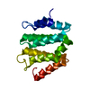
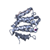
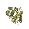
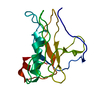
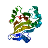
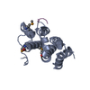
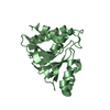
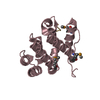
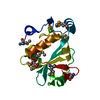
| Title | Complex of GGA3-VHS Domain and CI-MPR C-terminal Phosphopeptide | ||||||
 Components Components |
| ||||||
 Keywords Keywords | SIGNALING PROTEIN / VHS domain / Protein-phosphopeptide complex | ||||||
| Function / homology |  Function and homology information Function and homology informationpositive regulation of lysosomal protein catabolic process / Retrograde transport at the Trans-Golgi-Network / insulin-like growth factor receptor activity / clathrin coat / response to tetrachloromethane / retromer complex binding / insulin-like growth factor binding / protein transporter activity / insulin-like growth factor II binding / Golgi to plasma membrane transport ...positive regulation of lysosomal protein catabolic process / Retrograde transport at the Trans-Golgi-Network / insulin-like growth factor receptor activity / clathrin coat / response to tetrachloromethane / retromer complex binding / insulin-like growth factor binding / protein transporter activity / insulin-like growth factor II binding / Golgi to plasma membrane transport / trans-Golgi network transport vesicle / host-mediated activation of viral process / protein targeting to lysosome / MET receptor recycling / retinoic acid binding / endocytic recycling / Golgi to plasma membrane protein transport / protein localization to cell surface / TBC/RABGAPs / Golgi Associated Vesicle Biogenesis / nuclear envelope lumen / D-mannose binding / negative regulation of amyloid-beta formation / animal organ regeneration / post-embryonic development / response to retinoic acid / endocytic vesicle / G-protein alpha-subunit binding / transport vesicle / protein catabolic process / phosphatidylinositol binding / liver development / receptor-mediated endocytosis / secretory granule membrane / trans-Golgi network membrane / ubiquitin binding / trans-Golgi network / regulation of protein stability / clathrin-coated endocytic vesicle membrane / phosphoprotein binding / protein destabilization / intracellular protein transport / small GTPase binding / recycling endosome membrane / late endosome / Cargo recognition for clathrin-mediated endocytosis / Clathrin-mediated endocytosis / signaling receptor activity / early endosome membrane / spermatogenesis / early endosome / lysosome / endosome / endosome membrane / positive regulation of apoptotic process / G protein-coupled receptor signaling pathway / Amyloid fiber formation / Golgi membrane / focal adhesion / Neutrophil degranulation / protein-containing complex binding / perinuclear region of cytoplasm / Golgi apparatus / enzyme binding / cell surface / signal transduction / protein-containing complex / extracellular exosome / membrane / identical protein binding / plasma membrane Similarity search - Function | ||||||
| Biological species |  Homo sapiens (human) Homo sapiens (human) | ||||||
| Method |  X-RAY DIFFRACTION / FOURIER SYNTHESIS / Resolution: 2.3 Å X-RAY DIFFRACTION / FOURIER SYNTHESIS / Resolution: 2.3 Å | ||||||
 Authors Authors | Kato, Y. / Misra, S. / Puertollano, R. / Hurley, J.H. / Bonifacino, J.S. | ||||||
 Citation Citation | Journal: Nat.Struct.Biol. / Year: 2002 Title: Phosphoregulation of sorting signal-VHS domain interactions by a direct electrostatic mechanism. Authors: Kato, Y. / Misra, S. / Puertollano, R. / Hurley, J.H. / Bonifacino, J.S. | ||||||
| History |
|
- Structure visualization
| Structure viewer | Molecule: MolmilJmol/JSmol |
|---|
- Downloads & links
-Download
| PDBx/mmCIF format | 1lf8.cif.gz | 145 KB | Display | PDBx/mmCIF format |
|---|---|---|---|---|
| PDB format | pdb1lf8.ent.gz | 115.9 KB | Display | PDB format |
| PDBx/mmJSON format | 1lf8.json.gz | Tree view | PDBx/mmJSON format | |
| Others |  Other downloads Other downloads |
-Validation report
| Arichive directory | https://data.pdbj.org/pub/pdb/validation_reports/lf/1lf8ftp://data.pdbj.org/pub/pdb/validation_reports/lf/1lf8 | HTTPS FTP |
|---|
-Related structure data
| Related structure data |  1juqS S: Starting model for refinement |
|---|---|
| Similar structure data |


-Links
 PDBj
PDBj


- Assembly
| Deposited unit | 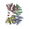
| ||||||||
|---|---|---|---|---|---|---|---|---|---|
| 1 | 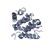
| ||||||||
| 2 | 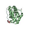
| ||||||||
| 3 | 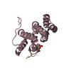
| ||||||||
| 4 | 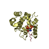
| ||||||||
| Unit cell |
| ||||||||
| Details | Each VHS domain/phosphopeptide complex corresponds to a single biological unit. There are the equivalent of 4 biological complexes per asymmetric unit. The apparent VHS domain dimer is likely a crystallization artifact. |
-Components
| #1: Protein | Mass: 19482.451 Da / Num. of mol.: 4 / Fragment: VHS domain (residues 1-166) Source method: isolated from a genetically manipulated source Source: (gene. exp.) Homo sapiens (human) / Plasmid: pHis-parallel2 / Species (production host): Escherichia coli / Production host:  #2: Protein/peptide | Mass: 1537.478 Da / Num. of mol.: 4 / Fragment: C-terminus (residues 2480-2491) / Source method: obtained synthetically Details: The peptide was chemically synthesized. The sequence of the peptide is naturally found in Homo sapiens (Human). References: UniProt: P11717 #3: Water | ChemComp-HOH / |  Mass: 18.015 Da / Num. of mol.: 300 / Source method: isolated from a natural source / Formula: H2O Mass: 18.015 Da / Num. of mol.: 300 / Source method: isolated from a natural source / Formula: H2OHas protein modification | Y | |
|---|
-Experimental details
-Experiment
| Experiment | Method: X-RAY DIFFRACTION / Number of used crystals: 1 |
|---|
- Sample preparation
| Crystal | Density Matthews: 2.61 Å3/Da / Density % sol: 52.8 % | |||||||||||||||||||||||||||||||||||||||||||||||||
|---|---|---|---|---|---|---|---|---|---|---|---|---|---|---|---|---|---|---|---|---|---|---|---|---|---|---|---|---|---|---|---|---|---|---|---|---|---|---|---|---|---|---|---|---|---|---|---|---|---|---|
| Crystal grow | Temperature: 293 K / Method: vapor diffusion, hanging drop Details: 100mM CAPS 10.2-11.0, 200 mM Lithium Sulfate, 1.3-2M Sodium/Potassium phosphate, VAPOR DIFFUSION, HANGING DROP, temperature 293K | |||||||||||||||||||||||||||||||||||||||||||||||||
| Crystal grow | *PLUS Temperature: 20 ℃ / pH: 8 | |||||||||||||||||||||||||||||||||||||||||||||||||
| Components of the solutions | *PLUS
|
-Data collection
| Diffraction | Mean temperature: 95 K |
|---|---|
| Diffraction source | Source: ROTATING ANODE / Type: RIGAKU RU200 / Wavelength: 1.5418 Å |
| Detector | Type: RIGAKU RAXIS IV / Detector: IMAGE PLATE / Date: Oct 25, 2001 / Details: Osmic Mirrors |
| Radiation | Protocol: SINGLE WAVELENGTH / Monochromatic (M) / Laue (L): M / Scattering type: x-ray |
| Radiation wavelength | Wavelength: 1.5418 Å / Relative weight: 1 |
| Reflection | Resolution: 2.3→100 Å / Num. obs: 37720 / % possible obs: 96.1 % / Observed criterion σ(I): -3 / Redundancy: 6.1 % / Biso Wilson estimate: 31.5 Å2 / Rmerge(I) obs: 0.062 / Net I/σ(I): 20.5 |
| Reflection shell | Resolution: 2.3→2.38 Å / Rmerge(I) obs: 0.356 / Mean I/σ(I) obs: 4.4 / Num. unique all: 3550 / % possible all: 91.5 |
| Reflection | *PLUS Highest resolution: 2.3 Å / Lowest resolution: 50 Å / Rmerge(I) obs: 0.062 |
| Reflection shell | *PLUS % possible obs: 91.5 % / Rmerge(I) obs: 0.356 |
- Processing
| Software |
| ||||||||||||||||||||||||||||||||||||
|---|---|---|---|---|---|---|---|---|---|---|---|---|---|---|---|---|---|---|---|---|---|---|---|---|---|---|---|---|---|---|---|---|---|---|---|---|---|
| Refinement | Method to determine structure: FOURIER SYNTHESIS Starting model: PDB ENTRY 1JUQ Resolution: 2.3→89.83 Å / Rfactor Rfree error: 0.004 / Data cutoff high absF: 713707.58 / Data cutoff low absF: 0 / Isotropic thermal model: RESTRAINED / Cross valid method: THROUGHOUT / σ(F): 0 / Stereochemistry target values: Engh & Huber
| ||||||||||||||||||||||||||||||||||||
| Solvent computation | Solvent model: FLAT MODEL / Bsol: 42.6607 Å2 / ksol: 0.338167 e/Å3 | ||||||||||||||||||||||||||||||||||||
| Displacement parameters | Biso mean: 49.8 Å2
| ||||||||||||||||||||||||||||||||||||
| Refine analyze |
| ||||||||||||||||||||||||||||||||||||
| Refinement step | Cycle: LAST / Resolution: 2.3→89.83 Å
| ||||||||||||||||||||||||||||||||||||
| Refine LS restraints |
| ||||||||||||||||||||||||||||||||||||
| LS refinement shell | Resolution: 2.3→2.44 Å / Rfactor Rfree error: 0.014 / Total num. of bins used: 6
| ||||||||||||||||||||||||||||||||||||
| Xplor file |
| ||||||||||||||||||||||||||||||||||||
| Refinement | *PLUS Highest resolution: 2.3 Å / Lowest resolution: 50 Å / % reflection Rfree: 10 % / Rfactor Rfree: 0.255 / Rfactor Rwork: 0.211 | ||||||||||||||||||||||||||||||||||||
| Solvent computation | *PLUS | ||||||||||||||||||||||||||||||||||||
| Displacement parameters | *PLUS | ||||||||||||||||||||||||||||||||||||
| Refine LS restraints | *PLUS
| ||||||||||||||||||||||||||||||||||||
| LS refinement shell | *PLUS Rfactor Rfree: 0.322 / Rfactor Rwork: 0.264 |
