[English] 日本語
Yorodumi
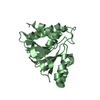
- PDB-1juq: GGA3 VHS domain complexed with C-terminal peptide from cation-dep... -

+
Open data


ID or keywords:

Loading...

-
Basic information

Entry
Database: PDB / ID: 1juq
TitleGGA3 VHS domain complexed with C-terminal peptide from cation-dependent Mannose 6-phosphate receptor
Components
  • ADP-RIBOSYLATION FACTOR BINDING PROTEIN GGA3
  • Cation-dependent mannose-6-phosphate receptor
KeywordsSIGNALING PROTEIN / protein-peptide complex / VHS domain / dxxll sorting signal
Function / homology
Function and homology information


secretion of lysosomal enzymes / Retrograde transport at the Trans-Golgi-Network / retromer complex binding / clathrin-coated vesicle membrane / Golgi to plasma membrane protein transport / protein targeting to lysosome / Golgi to plasma membrane transport / membrane organization / endocytic recycling / MET receptor recycling ...secretion of lysosomal enzymes / Retrograde transport at the Trans-Golgi-Network / retromer complex binding / clathrin-coated vesicle membrane / Golgi to plasma membrane protein transport / protein targeting to lysosome / Golgi to plasma membrane transport / membrane organization / endocytic recycling / MET receptor recycling / protein localization to cell surface / TBC/RABGAPs / lysosomal transport / Lysosome Vesicle Biogenesis / endosome to lysosome transport / negative regulation of amyloid-beta formation / plasma membrane => GO:0005886 / transport vesicle / receptor-mediated endocytosis / phosphatidylinositol binding / trans-Golgi network membrane / ubiquitin binding / intracellular protein transport / protein catabolic process / protein destabilization / regulation of protein stability / trans-Golgi network / small GTPase binding / recycling endosome membrane / positive regulation of protein catabolic process / transmembrane signaling receptor activity / late endosome / Cargo recognition for clathrin-mediated endocytosis / Clathrin-mediated endocytosis / early endosome membrane / lysosome / endosome membrane / endosome / Amyloid fiber formation / lysosomal membrane / protein domain specific binding / protein-containing complex binding / perinuclear region of cytoplasm / Golgi apparatus / protein-containing complex / membrane / plasma membrane
Similarity search - Function
GGA3, GAT domain / : / Cation-dependent mannose-6-phosphate receptor / Mannose-6-phosphate receptor / Mannose-6-phosphate receptor / ADP-ribosylation factor-binding protein GGA3 / N-terminal extension of GAT domain / N-terminal extension of GAT domain / GAT domain / GAT domain superfamily ...GGA3, GAT domain / : / Cation-dependent mannose-6-phosphate receptor / Mannose-6-phosphate receptor / Mannose-6-phosphate receptor / ADP-ribosylation factor-binding protein GGA3 / N-terminal extension of GAT domain / N-terminal extension of GAT domain / GAT domain / GAT domain superfamily / GAT domain / GAT domain profile. / VHS domain / VHS domain / VHS domain profile. / Domain present in VPS-27, Hrs and STAM / MRH domain profile. / Gamma-adaptin ear (GAE) domain / Gamma-adaptin ear (GAE) domain profile. / Mannose-6-phosphate receptor binding domain superfamily / Serine Threonine Protein Phosphatase 5, Tetratricopeptide repeat - #90 / Clathrin adaptor, alpha/beta/gamma-adaptin, appendage, Ig-like subdomain / Adaptin C-terminal domain / Adaptin C-terminal domain / ENTH/VHS / Clathrin adaptor, appendage, Ig-like subdomain superfamily / Serine Threonine Protein Phosphatase 5, Tetratricopeptide repeat / Alpha Horseshoe / Mainly Alpha
Similarity search - Domain/homology
Cation-dependent mannose-6-phosphate receptor / ADP-ribosylation factor-binding protein GGA3
Similarity search - Component
Biological speciesHomo sapiens (human)
MethodX-RAY DIFFRACTION / SYNCHROTRON / MAD/FOURIER SYNTHESIS / Resolution: 2.2 Å
AuthorsMisra, S. / Puertollano, R. / Bonifacino, J.S. / Hurley, J.H.
CitationJournal: Nature / Year: 2002
Title: Structural basis for acidic-cluster-dileucine sorting-signal recognition by VHS domains.
Authors: Misra, S. / Puertollano, R. / Kato, Y. / Bonifacino, J.S. / Hurley, J.H.
History
DepositionAug 26, 2001Deposition site: RCSB / Processing site: RCSB
Revision 1.0Feb 27, 2002Provider: repository / Type: Initial release
Revision 1.1Apr 27, 2008Group: Version format compliance
Revision 1.2Jul 13, 2011Group: Version format compliance
Revision 1.3Oct 12, 2016Group: Advisory
Remark 300
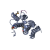
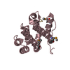
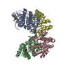
BIOMOLECULE: 1, 2, 3, 4 THIS ENTRY CONTAINS THE CRYSTALLOGRAPHIC ASYMMETRIC UNIT WHICH CONSISTS OF ...BIOMOLECULE: 1, 2, 3, 4 THIS ENTRY CONTAINS THE CRYSTALLOGRAPHIC ASYMMETRIC UNIT WHICH CONSISTS OF 8 CHAIN(S). SEE REMARK 350 FOR INFORMATION ON GENERATING THE BIOLOGICAL MOLECULE(S). The authors have gel filtration and analytical ultracentrifugation data that suggest that the asymmetric unit contains 4 biological units (chains A/E, B/F, C/G, D/H). The authors think the apparent dimer in the crystal is due to crystallization.

-
Structure visualization

Structure viewerMolecule:
MolmilJmol/JSmol

Downloads & links

-
Assembly

Deposited unit
A: ADP-RIBOSYLATION FACTOR BINDING PROTEIN GGA3
E: Cation-dependent mannose-6-phosphate receptor
B: ADP-RIBOSYLATION FACTOR BINDING PROTEIN GGA3
F: Cation-dependent mannose-6-phosphate receptor
C: ADP-RIBOSYLATION FACTOR BINDING PROTEIN GGA3
G: Cation-dependent mannose-6-phosphate receptor
D: ADP-RIBOSYLATION FACTOR BINDING PROTEIN GGA3
H: Cation-dependent mannose-6-phosphate receptor


Theoretical massNumber of molelcules
Total (without water)85,2748
Polymers85,2748
Non-polymers00
Water3,153175
1
A: ADP-RIBOSYLATION FACTOR BINDING PROTEIN GGA3
E: Cation-dependent mannose-6-phosphate receptor


Theoretical massNumber of molelcules
Total (without water)21,3192
Polymers21,3192
Non-polymers00
Water362
TypeNameSymmetry operationNumber
identity operation1_555x,y,z1
Buried area750 Å2
ΔGint-6 kcal/mol
Surface area8620 Å2
MethodPISA
2
B: ADP-RIBOSYLATION FACTOR BINDING PROTEIN GGA3
F: Cation-dependent mannose-6-phosphate receptor


Theoretical massNumber of molelcules
Total (without water)21,3192
Polymers21,3192
Non-polymers00
Water181
TypeNameSymmetry operationNumber
identity operation1_555x,y,z1
Buried area990 Å2
ΔGint-8 kcal/mol
Surface area8880 Å2
MethodPISA
3
C: ADP-RIBOSYLATION FACTOR BINDING PROTEIN GGA3
G: Cation-dependent mannose-6-phosphate receptor


Theoretical massNumber of molelcules
Total (without water)21,3192
Polymers21,3192
Non-polymers00
Water181
TypeNameSymmetry operationNumber
identity operation1_555x,y,z1
4
D: ADP-RIBOSYLATION FACTOR BINDING PROTEIN GGA3
H: Cation-dependent mannose-6-phosphate receptor


Theoretical massNumber of molelcules
Total (without water)21,3192
Polymers21,3192
Non-polymers00
Water362
TypeNameSymmetry operationNumber
identity operation1_555x,y,z1
Buried area940 Å2
ΔGint-7 kcal/mol
Surface area9180 Å2
MethodPISA
5
A: ADP-RIBOSYLATION FACTOR BINDING PROTEIN GGA3
E: Cation-dependent mannose-6-phosphate receptor
D: ADP-RIBOSYLATION FACTOR BINDING PROTEIN GGA3
H: Cation-dependent mannose-6-phosphate receptor

B: ADP-RIBOSYLATION FACTOR BINDING PROTEIN GGA3
F: Cation-dependent mannose-6-phosphate receptor
C: ADP-RIBOSYLATION FACTOR BINDING PROTEIN GGA3
G: Cation-dependent mannose-6-phosphate receptor


Theoretical massNumber of molelcules
Total (without water)85,2748
Polymers85,2748
Non-polymers00
Water1086
TypeNameSymmetry operationNumber
identity operation1_555x,y,z1
crystal symmetry operation6_555-x+1/2,-y+1/2,z+1/21
Buried area8240 Å2
ΔGint-49 kcal/mol
Surface area30310 Å2
MethodPISA
6
A: ADP-RIBOSYLATION FACTOR BINDING PROTEIN GGA3
E: Cation-dependent mannose-6-phosphate receptor
D: ADP-RIBOSYLATION FACTOR BINDING PROTEIN GGA3

B: ADP-RIBOSYLATION FACTOR BINDING PROTEIN GGA3
C: ADP-RIBOSYLATION FACTOR BINDING PROTEIN GGA3
G: Cation-dependent mannose-6-phosphate receptor


Theoretical massNumber of molelcules
Total (without water)82,0716
Polymers82,0716
Non-polymers00
Water905
TypeNameSymmetry operationNumber
identity operation1_555x,y,z1
crystal symmetry operation6_555-x+1/2,-y+1/2,z+1/21
Buried area6310 Å2
ΔGint-34 kcal/mol
Surface area29730 Å2
MethodPISA
Unit cell
Length a, b, c (Å)123.640, 131.350, 108.140
Angle α, β, γ (deg.)90.00, 90.00, 90.00
Int Tables number20
Space group name H-MC2221

-
Components

#1: Protein
ADP-RIBOSYLATION FACTOR BINDING PROTEIN GGA3


Mass: 19716.924 Da / Num. of mol.: 4 / Fragment: VHS domain
Source method: isolated from a genetically manipulated source
Source: (gene. exp.) Homo sapiens (human) / Gene: GGA3 / Plasmid: pHis-parallel2 / Species (production host): Escherichia coli / Production host: Escherichia coli BL21(DE3) (bacteria) / Strain (production host): BL21(DE3) / References: UniProt: Q9NZ52
#2: Protein/peptide
Cation-dependent mannose-6-phosphate receptor /


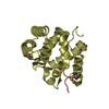
Mass: 1601.691 Da / Num. of mol.: 4 / Fragment: C-terminal peptide / Source method: obtained synthetically
Details: The peptide was chemically synthesized. The sequence of the peptide is naturally found in Homo sapiens (human).
References: UniProt: P20645
#3: Water ChemComp-HOH / water / Water


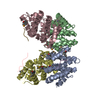
Mass: 18.015 Da / Num. of mol.: 175 / Source method: isolated from a natural source / Formula: H2O

-
Experimental details

-
Experiment

ExperimentMethod: X-RAY DIFFRACTION / Number of used crystals: 2

-
Sample preparation

CrystalDensity Matthews: 2.57 Å3/Da / Density % sol: 52.19 %
Crystal growTemperature: 293 K / Method: vapor diffusion, hanging drop
Details: Sodium/Potassium phosphate, Lithium Sulfate, CAPS, VAPOR DIFFUSION, HANGING DROP, temperature 293K
Crystal grow
*PLUS
PH range low: 11 / PH range high: 10.2
Components of the solutions
*PLUS
IDConc.Common nameCrystal-IDSol-IDDetailsChemical formula
112 mg/mlprotein1drop
21.5 mMpeptide1drop
3100 mMCAPS1reservoirpH10.2-11.0
4200 mM1reservoirLi2SO4
51.33-2 Msodium potassium phospahte1reservoir

-
Data collection

Diffraction
IDMean temperature (K)Crystal-ID
1951
21001
Diffraction source
SourceSiteBeamlineTypeIDWavelength (Å)
ROTATING ANODERIGAKU11.5418
SYNCHROTRONNSLS X9B20.97900,0.97946,0.96859
Detector
TypeIDDetectorDateDetails
RIGAKU RAXIS IV1IMAGE PLATEJun 23, 2001mirrors
ADSC QUANTUM 42CCDJun 3, 2001
Radiation
IDMonochromatorProtocolMonochromatic (M) / Laue (L)Scattering typeWavelength-ID
1GraphiteSINGLE WAVELENGTHMx-ray1
2SAGITALLY FOCUSED Si(111)MADMx-ray1
Radiation wavelength
IDWavelength (Å)Relative weight
11.54181
20.9791
30.979461
40.968591
ReflectionResolution: 2.2→100 Å / Num. obs: 43908 / % possible obs: 97.6 % / Observed criterion σ(F): 2 / Redundancy: 3.57 % / Biso Wilson estimate: 28.5 Å2 / Rmerge(I) obs: 0.051 / Net I/σ(I): 19.5
Reflection shellResolution: 2.2→2.28 Å / Redundancy: 2.8 % / Rmerge(I) obs: 0.282 / Mean I/σ(I) obs: 3.97 / Num. unique all: 3823 / % possible all: 86.7
Reflection shell
*PLUS
% possible obs: 86.7 % / Num. unique obs: 3823 / Mean I/σ(I) obs: 4

-
Processing

Software
NameVersionClassification
SOLVEphasing
CNS1refinement
DENZOdata reduction
SCALEPACKdata scaling
RefinementMethod to determine structure: MAD/FOURIER SYNTHESIS / Resolution: 2.2→90.03 Å / Rfactor Rfree error: 0.004 / Data cutoff high absF: 628452.57 / Data cutoff low absF: 0 / Isotropic thermal model: RESTRAINED / Cross valid method: THROUGHOUT / σ(F): 0 / Stereochemistry target values: Engh & Huber
RfactorNum. reflection% reflectionSelection details
Rfree0.254 4269 10.1 %RANDOM
Rwork0.221 ---
obs-42356 94.2 %-
Solvent computationSolvent model: FLAT MODEL / Bsol: 49.3598 Å2 / ksol: 0.385002 e/Å3
Displacement parametersBiso mean: 45.9 Å2
Baniso -1Baniso -2Baniso -3
1--5.89 Å20 Å20 Å2
2--13.22 Å20 Å2
3----7.33 Å2
Refine analyze
FreeObs
Luzzati coordinate error0.33 Å0.27 Å
Luzzati d res low-5 Å
Luzzati sigma a0.23 Å0.2 Å
Refinement stepCycle: LAST / Resolution: 2.2→90.03 Å
ProteinNucleic acidLigandSolventTotal
Num. atoms5214 0 0 175 5389
Refine LS restraints
Refine-IDTypeDev idealDev ideal target
X-RAY DIFFRACTIONc_bond_d0.006
X-RAY DIFFRACTIONc_angle_deg1.2
X-RAY DIFFRACTIONc_dihedral_angle_d18.7
X-RAY DIFFRACTIONc_improper_angle_d0.79
X-RAY DIFFRACTIONc_mcbond_it1.441.5
X-RAY DIFFRACTIONc_mcangle_it2.312
X-RAY DIFFRACTIONc_scbond_it2.342
X-RAY DIFFRACTIONc_scangle_it3.462.5
LS refinement shellResolution: 2.2→2.34 Å / Rfactor Rfree error: 0.012 / Total num. of bins used: 6
RfactorNum. reflection% reflection
Rfree0.284 597 9.9 %
Rwork0.255 5439 -
obs--81.7 %
Xplor file
Refine-IDSerial noParam fileTopol file
X-RAY DIFFRACTION1PROTEIN_REP.PARAMPROTEIN.TOP
X-RAY DIFFRACTION2WATER_REP.PARAMWATER.TOP
X-RAY DIFFRACTION3ION.PARAMION.TOP
Software
*PLUS
Name: CNS / Version: 1 / Classification: refinement
Refinement
*PLUS
Highest resolution: 2.2 Å / Lowest resolution: 90 Å / σ(F): 0 / % reflection Rfree: 10 % / Rfactor obs: 0.224 / Rfactor Rfree: 0.256
Solvent computation
*PLUS
Displacement parameters
*PLUS
Biso mean: 45.9 Å2
Refine LS restraints
*PLUS
Refine-IDTypeDev idealDev ideal target
X-RAY DIFFRACTIONc_dihedral_angle_d
X-RAY DIFFRACTIONc_dihedral_angle_deg18.7
X-RAY DIFFRACTIONc_improper_angle_d
X-RAY DIFFRACTIONc_improper_angle_deg0.79
X-RAY DIFFRACTIONc_mcbond_it1.441.5
X-RAY DIFFRACTIONc_scbond_it2.342
X-RAY DIFFRACTIONc_mcangle_it2.312
X-RAY DIFFRACTIONc_scangle_it3.462.5
LS refinement shell
*PLUS
Rfactor Rfree: 0.288 / % reflection Rfree: 9.9 % / Rfactor Rwork: 0.255 / Rfactor obs: 0.258

+
About Yorodumi

-
News

-
Feb 9, 2022. New format data for meta-information of EMDB entries

New format data for meta-information of EMDB entries

  • Version 3 of the EMDB header file is now the official format.
  • The previous official version 1.9 will be removed from the archive.

Related info.:EMDB header

External links:wwPDB to switch to version 3 of the EMDB data model

-
Aug 12, 2020. Covid-19 info

Covid-19 info

URL: https://pdbj.org/emnavi/covid19.php

New page: Covid-19 featured information page in EM Navigator.

Related info.:Covid-19 info / Mar 5, 2020. Novel coronavirus structure data

+
Mar 5, 2020. Novel coronavirus structure data

Novel coronavirus structure data

Related info.:Yorodumi Speices / Aug 12, 2020. Covid-19 info

External links:COVID-19 featured content - PDBj / Molecule of the Month (242):Coronavirus Proteases

+
Jan 31, 2019. EMDB accession codes are about to change! (news from PDBe EMDB page)

EMDB accession codes are about to change! (news from PDBe EMDB page)

  • The allocation of 4 digits for EMDB accession codes will soon come to an end. Whilst these codes will remain in use, new EMDB accession codes will include an additional digit and will expand incrementally as the available range of codes is exhausted. The current 4-digit format prefixed with “EMD-” (i.e. EMD-XXXX) will advance to a 5-digit format (i.e. EMD-XXXXX), and so on. It is currently estimated that the 4-digit codes will be depleted around Spring 2019, at which point the 5-digit format will come into force.
  • The EM Navigator/Yorodumi systems omit the EMD- prefix.

Related info.:Q: What is EMD? / ID/Accession-code notation in Yorodumi/EM Navigator

External links:EMDB Accession Codes are Changing Soon! / Contact to PDBj

+
Jul 12, 2017. Major update of PDB

Major update of PDB

  • wwPDB released updated PDB data conforming to the new PDBx/mmCIF dictionary.
  • This is a major update changing the version number from 4 to 5, and with Remediation, in which all the entries are updated.
  • In this update, many items about electron microscopy experimental information are reorganized (e.g. em_software).
  • Now, EM Navigator and Yorodumi are based on the updated data.

External links:wwPDB Remediation / Enriched Model Files Conforming to OneDep Data Standards Now Available in the PDB FTP Archive

-
Yorodumi

Thousand views of thousand structures

  • Yorodumi is a browser for structure data from EMDB, PDB, SASBDB, etc.
  • This page is also the successor to EM Navigator detail page, and also detail information page/front-end page for Omokage search.
  • The word "yorodu" (or yorozu) is an old Japanese word meaning "ten thousand". "mi" (miru) is to see.

Related info.:EMDB / PDB / SASBDB / Comparison of 3 databanks / Yorodumi Search / Aug 31, 2016. New EM Navigator & Yorodumi / Yorodumi Papers / Jmol/JSmol / Function and homology information / Changes in new EM Navigator and Yorodumi

Read more