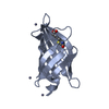
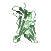
 Movie
Movie Controller


+ Open data

- Basic information
| Entry | Database: PDB / ID: 1e6f | ||||||
|---|---|---|---|---|---|---|---|
| Title | Human MIR-receptor, repeat 11 | ||||||
 Components Components | CATION-INDEPENDENT MANNOSE-6-PHOSPHATE RECEPTOR | ||||||
 Keywords Keywords | RECEPTOR / MIR-RECEPTOR / IGF-II RECEPTOR / TRANSPORT / GLYCOPROTEIN | ||||||
| Function / homology |  Function and homology information Function and homology informationRetrograde transport at the Trans-Golgi-Network / insulin-like growth factor receptor activity / clathrin coat / response to tetrachloromethane / retromer complex binding / insulin-like growth factor binding / insulin-like growth factor II binding / trans-Golgi network transport vesicle / host-mediated activation of viral process / protein targeting to lysosome ...Retrograde transport at the Trans-Golgi-Network / insulin-like growth factor receptor activity / clathrin coat / response to tetrachloromethane / retromer complex binding / insulin-like growth factor binding / insulin-like growth factor II binding / trans-Golgi network transport vesicle / host-mediated activation of viral process / protein targeting to lysosome / retinoic acid binding / nuclear envelope lumen / Golgi Associated Vesicle Biogenesis / D-mannose binding / animal organ regeneration / response to retinoic acid / endocytic vesicle / G-protein alpha-subunit binding / transport vesicle / post-embryonic development / receptor-mediated endocytosis / secretory granule membrane / liver development / trans-Golgi network membrane / trans-Golgi network / clathrin-coated endocytic vesicle membrane / phosphoprotein binding / late endosome / Cargo recognition for clathrin-mediated endocytosis / Clathrin-mediated endocytosis / signaling receptor activity / spermatogenesis / early endosome / endosome / endosome membrane / positive regulation of apoptotic process / G protein-coupled receptor signaling pathway / Golgi membrane / focal adhesion / Neutrophil degranulation / perinuclear region of cytoplasm / Golgi apparatus / enzyme binding / cell surface / signal transduction / extracellular exosome / membrane / identical protein binding / plasma membrane Similarity search - Function | ||||||
| Biological species |  HOMO SAPIENS (human) HOMO SAPIENS (human) | ||||||
| Method |  X-RAY DIFFRACTION / SYNCHROTRON / SIRAS / Resolution: 1.75 Å X-RAY DIFFRACTION / SYNCHROTRON / SIRAS / Resolution: 1.75 Å | ||||||
 Authors Authors | Von Buelow, R. / Rajashankar, K.R. / Dauter, M. / Dauter, Z. / Grimme, S. / Schmidt, B. / Von Figura, K. / Uson, I. | ||||||
 Citation Citation | Journal: Acta Crystallogr.,Sect.D / Year: 2003 Title: Locating the Anomalous Scatterer Substructures in Halide and Sulfur Phasing Authors: Uson, I. / Schmidt, B. / Von Buelow, R. / Grimme, S. / Von Figura, K. / Dauter, M. / Rajashankar, K.R. / Dauter, Z. / Sheldrick, G.M. | ||||||
| History |
|
- Structure visualization
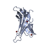
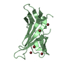
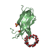
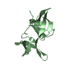
| Structure viewer | Molecule: MolmilJmol/JSmol |
|---|
- Downloads & links
-Download
| PDBx/mmCIF format | 1e6f.cif.gz | 65.1 KB | Display | PDBx/mmCIF format |
|---|---|---|---|---|
| PDB format | pdb1e6f.ent.gz | 47.7 KB | Display | PDB format |
| PDBx/mmJSON format | 1e6f.json.gz | Tree view | PDBx/mmJSON format | |
| Others |  Other downloads Other downloads |
-Validation report
| Arichive directory | https://data.pdbj.org/pub/pdb/validation_reports/e6/1e6fftp://data.pdbj.org/pub/pdb/validation_reports/e6/1e6f | HTTPS FTP |
|---|
-Related structure data


-Links
 PDBj
PDBj


- Assembly
| Deposited unit | 
| ||||||||
|---|---|---|---|---|---|---|---|---|---|
| 1 | 
| ||||||||
| 2 | 
| ||||||||
| Unit cell |
| ||||||||
| Noncrystallographic symmetry (NCS) | NCS oper: (Code: given Matrix: (0.02057, -0.99979, -0.00014), Vector: |
-Components
| #1: Protein | Mass: 15564.672 Da / Num. of mol.: 2 Fragment: IGF-II-BINDING DOMAIN, REPEAT 11, RESIDUES 1508-1650 Source method: isolated from a genetically manipulated source Source: (gene. exp.) HOMO SAPIENS (human) / Cell: FIBROBLAST / Cell line: BHK-21 / Cellular location: LYSOSOME / Organ: KIDNEY / Production host:  CRICETINAE GEN. SP. (mammal) / References: UniProt: P11717 CRICETINAE GEN. SP. (mammal) / References: UniProt: P11717#2: Water | ChemComp-HOH / |  Mass: 18.015 Da / Num. of mol.: 118 / Source method: isolated from a natural source / Formula: H2O Mass: 18.015 Da / Num. of mol.: 118 / Source method: isolated from a natural source / Formula: H2OCompound details | TRANSPORT OF PHOSPHORYLATED LYSOSOMAL ENZYMES FROM THE GOLGI COMPLEX AND THE CELL SURFACE TO ...TRANSPORT OF PHOSPHORYL | Has protein modification | Y | |
|---|
-Experimental details
-Experiment
| Experiment | Method: X-RAY DIFFRACTION / Number of used crystals: 1 |
|---|
- Sample preparation
| Crystal | Density Matthews: 2.17 Å3/Da / Density % sol: 40 % | ||||||||||||||||||||||||||||||||||||||||||
|---|---|---|---|---|---|---|---|---|---|---|---|---|---|---|---|---|---|---|---|---|---|---|---|---|---|---|---|---|---|---|---|---|---|---|---|---|---|---|---|---|---|---|---|
| Crystal grow | Method: vapor diffusion, hanging drop / pH: 5.7 Details: PRECIPITANT: 0.2 M AMMONIUM ACETATE, 0.1 M CACODYLATE PH 5 28% PEG 4000. PROTEIN SOLUTION: 8 MG/ML IN 10 MM TRIS-HCL PH7.5, 150 MM N VAPOUR DIFFUSION, HANGING DROPS,1:1 RATIO. | ||||||||||||||||||||||||||||||||||||||||||
| Crystal | *PLUS Density % sol: 35 % | ||||||||||||||||||||||||||||||||||||||||||
| Crystal grow | *PLUS Temperature: 288 K / pH: 7.5 / Method: vapor diffusion, hanging drop | ||||||||||||||||||||||||||||||||||||||||||
| Components of the solutions | *PLUS
|
-Data collection
| Diffraction | Mean temperature: 100 K |
|---|---|
| Diffraction source | Source: SYNCHROTRON / Site: EMBL/DESY, HAMBURG  / Beamline: X11 / Wavelength: 0.9057 / Beamline: X11 / Wavelength: 0.9057 |
| Detector | Type: MARRESEARCH / Detector: IMAGE PLATE / Date: Aug 15, 1999 / Details: BENT CRYSTALS |
| Radiation | Monochromator: SILICON / Protocol: SINGLE WAVELENGTH / Monochromatic (M) / Laue (L): M / Scattering type: x-ray |
| Radiation wavelength | Wavelength: 0.9057 Å / Relative weight: 1 |
| Reflection | Resolution: 1.753→8 Å / Num. obs: 29383 / % possible obs: 99.1 % / Redundancy: 15 % / Rmerge(I) obs: 0.025 / Net I/σ(I): 40.8 |
| Reflection shell | Resolution: 1.75→1.85 Å / Redundancy: 5 % / Rmerge(I) obs: 0.147 / Mean I/σ(I) obs: 9.9 / % possible all: 97.3 |
| Reflection | *PLUS Lowest resolution: 8 Å / Redundancy: 8.3 % |
| Reflection shell | *PLUS % possible obs: 97.4 % / Rmerge(I) obs: 0.089 |
- Processing
| Software |
| |||||||||||||||||||||||||||||||||
|---|---|---|---|---|---|---|---|---|---|---|---|---|---|---|---|---|---|---|---|---|---|---|---|---|---|---|---|---|---|---|---|---|---|---|
| Refinement | Method to determine structure: SIRAS / Resolution: 1.75→8 Å / Num. parameters: 8499 / Num. restraintsaints: 10697 / Cross valid method: FREE R-VALUE / σ(F): 0 / Stereochemistry target values: ENGH AND HUBER / Details: REFINED OCCUPANCY FOR ALTERNATIVE DISORDERED SITES
| |||||||||||||||||||||||||||||||||
| Solvent computation | Solvent model: MOEWS & KRETSINGER, J.MOL.BIOL.91(1973)201-2 | |||||||||||||||||||||||||||||||||
| Refine analyze | Num. disordered residues: 22 / Occupancy sum hydrogen: 0 / Occupancy sum non hydrogen: 2050.5 | |||||||||||||||||||||||||||||||||
| Refinement step | Cycle: LAST / Resolution: 1.75→8 Å
| |||||||||||||||||||||||||||||||||
| Refine LS restraints |
| |||||||||||||||||||||||||||||||||
| Software | *PLUS Name: SHELXL-97 / Classification: refinement | |||||||||||||||||||||||||||||||||
| Refinement | *PLUS Rfactor Rwork: 0.2209 | |||||||||||||||||||||||||||||||||
| Solvent computation | *PLUS | |||||||||||||||||||||||||||||||||
| Displacement parameters | *PLUS |
