 Movie
Movie Controller

[English] 日本語
 Yorodumi
- PDB-2v2l: Mutant (E53,56,57,60Q) recombinant horse spleen apoferritin cocry... -
+ Open data

- Basic information
| Entry | Database: PDB / ID: 2v2l | ||||||
|---|---|---|---|---|---|---|---|
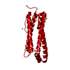
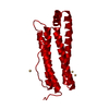
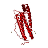
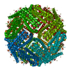
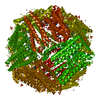
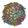
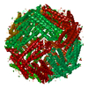
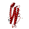
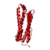
| Title | Mutant (E53,56,57,60Q) recombinant horse spleen apoferritin cocrystallized with haemin in acidic conditions | ||||||
 Components Components | FERRITIN LIGHT CHAIN | ||||||
 Keywords Keywords | METAL TRANSPORT / IRON / IRON STORAGE / METAL-BINDING | ||||||
| Function / homology |  Function and homology information Function and homology informationferritin complex / autolysosome / ferric iron binding / autophagosome / iron ion transport / ferrous iron binding / cytoplasmic vesicle / intracellular iron ion homeostasis / iron ion binding / cytoplasm Similarity search - Function | ||||||
| Biological species |  | ||||||
| Method |  X-RAY DIFFRACTION / SYNCHROTRON / MOLECULAR REPLACEMENT / Resolution: 1.9 Å X-RAY DIFFRACTION / SYNCHROTRON / MOLECULAR REPLACEMENT / Resolution: 1.9 Å | ||||||
 Authors Authors | De Val, N. / Declercq, J.P. | ||||||
 Citation Citation | Journal: J.Inorg.Biochem. / Year: 2012 Title: Structural Analysis of Haemin Demetallation by L-Chain Apoferritins Authors: De Val, N. / Declercq, J.P. / Lim, C.K. / Crichton, R.R. | ||||||
| History |
| ||||||
| Remark 650 | HELIX DETERMINATION METHOD: AUTHOR PROVIDED. |
- Structure visualization
| Structure viewer | Molecule: MolmilJmol/JSmol |
|---|
- Downloads & links
-Download
| PDBx/mmCIF format | 2v2l.cif.gz | 54.8 KB | Display | PDBx/mmCIF format |
|---|---|---|---|---|
| PDB format | pdb2v2l.ent.gz | 40 KB | Display | PDB format |
| PDBx/mmJSON format | 2v2l.json.gz | Tree view | PDBx/mmJSON format | |
| Others |  Other downloads Other downloads |
-Validation report
| Arichive directory | https://data.pdbj.org/pub/pdb/validation_reports/v2/2v2lftp://data.pdbj.org/pub/pdb/validation_reports/v2/2v2l | HTTPS FTP |
|---|
-Related structure data
| Related structure data | 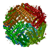 2v2iSC 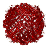 2v2jC 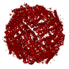 2v2mC  2v2nC  2v2oC  2v2pC  2v2rC 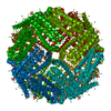 2v2sC  2w0oC S: Starting model for refinement C: citing same article ( |
|---|---|
| Similar structure data |


-Links
 PDBj
PDBj


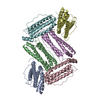
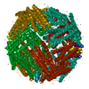
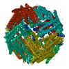
- Assembly
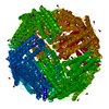
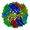
| Deposited unit | 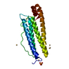
| |||||||||
|---|---|---|---|---|---|---|---|---|---|---|
| 1 | x 24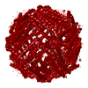
| |||||||||
| Unit cell |
| |||||||||
| Components on special symmetry positions |
|
-Components
| #1: Protein | Mass: 19852.445 Da / Num. of mol.: 1 / Mutation: YES Source method: isolated from a genetically manipulated source Source: (gene. exp.)  | ||||||||||
|---|---|---|---|---|---|---|---|---|---|---|---|
| #2: Chemical | ChemComp-CD /   Mass: 112.411 Da / Num. of mol.: 7 / Source method: obtained synthetically / Formula: Cd Mass: 112.411 Da / Num. of mol.: 7 / Source method: obtained synthetically / Formula: Cd#3: Chemical | ChemComp-SO4 / |   Mass: 96.063 Da / Num. of mol.: 1 / Source method: obtained synthetically / Formula: SO4 Mass: 96.063 Da / Num. of mol.: 1 / Source method: obtained synthetically / Formula: SO4#4: Chemical | ChemComp-GOL / |   Mass: 92.094 Da / Num. of mol.: 1 / Source method: obtained synthetically / Formula: C3H8O3 Mass: 92.094 Da / Num. of mol.: 1 / Source method: obtained synthetically / Formula: C3H8O3#5: Water | ChemComp-HOH / |  Mass: 18.015 Da / Num. of mol.: 174 / Source method: isolated from a natural source / Formula: H2O Mass: 18.015 Da / Num. of mol.: 174 / Source method: isolated from a natural source / Formula: H2OCompound details | ENGINEERED RESIDUE IN CHAIN A, GLU 53 TO GLN ENGINEERED RESIDUE IN CHAIN A, GLU 56 TO GLN ...ENGINEERED | Sequence details | MUTATIONS E53,56,57,60Q | |
-Experimental details
-Experiment
| Experiment | Method: X-RAY DIFFRACTION / Number of used crystals: 1 |
|---|
- Sample preparation
| Crystal | Density Matthews: 3.2 Å3/Da / Density % sol: 61.2 % / Description: NONE |
|---|---|
| Crystal grow | pH: 5.5 Details: RESERVOIR: CADMIUM SULFATE 0.13M, AMMONIUM SULFATE 0.8M, SODIUM ACETATE 0.1M PH 5.5, SODIUM AZIDE 0.003M. DROP: 2 UL PROTEIN AND 2 UL RESERVOIR |
-Data collection
| Diffraction | Mean temperature: 100 K |
|---|---|
| Diffraction source | Source: SYNCHROTRON / Site: ESRF  / Beamline: BM30A / Wavelength: 0.97991 / Beamline: BM30A / Wavelength: 0.97991 |
| Detector | Type: MARRESEARCH / Detector: CCD / Date: Nov 14, 2003 / Details: TWO MIRRORS ARE USED FOR VERTICAL FOCUSSING. |
| Radiation | Monochromator: FIRST CRYSTAL FLAT AND N2 COOLED. SECOND ONE SAGITALLY BENT. SI(111) OR SI(311) Protocol: SINGLE WAVELENGTH / Monochromatic (M) / Laue (L): M / Scattering type: x-ray |
| Radiation wavelength | Wavelength: 0.97991 Å / Relative weight: 1 |
| Reflection | Resolution: 1.9→20 Å / Num. obs: 21041 / % possible obs: 100 % / Observed criterion σ(I): 0 / Redundancy: 10.5 % / Rmerge(I) obs: 0.06 / Net I/σ(I): 37.1 |
| Reflection shell | Resolution: 1.9→1.94 Å / Redundancy: 7 % / Rmerge(I) obs: 0.49 / Mean I/σ(I) obs: 5.3 / % possible all: 99.9 |
- Processing
| Software |
| ||||||||||||||||||||||||||||||||||||||||||||||||||||||||||||||||||||||||||||||||||||||||||||||||||||||||||||||||||||||||||||||||||||||||||||||||||||||||||||||||||||||||||||||||||||||
|---|---|---|---|---|---|---|---|---|---|---|---|---|---|---|---|---|---|---|---|---|---|---|---|---|---|---|---|---|---|---|---|---|---|---|---|---|---|---|---|---|---|---|---|---|---|---|---|---|---|---|---|---|---|---|---|---|---|---|---|---|---|---|---|---|---|---|---|---|---|---|---|---|---|---|---|---|---|---|---|---|---|---|---|---|---|---|---|---|---|---|---|---|---|---|---|---|---|---|---|---|---|---|---|---|---|---|---|---|---|---|---|---|---|---|---|---|---|---|---|---|---|---|---|---|---|---|---|---|---|---|---|---|---|---|---|---|---|---|---|---|---|---|---|---|---|---|---|---|---|---|---|---|---|---|---|---|---|---|---|---|---|---|---|---|---|---|---|---|---|---|---|---|---|---|---|---|---|---|---|---|---|---|---|
| Refinement | Method to determine structure: MOLECULAR REPLACEMENT Starting model: PDB ENTRY 2V2I Resolution: 1.9→105.41 Å / Cor.coef. Fo:Fc: 0.951 / Cor.coef. Fo:Fc free: 0.939 / SU B: 2.388 / SU ML: 0.072 / Cross valid method: THROUGHOUT / ESU R: 0.125 / ESU R Free: 0.119 / Stereochemistry target values: MAXIMUM LIKELIHOOD / Details: HYDROGENS HAVE BEEN ADDED IN THE RIDING POSITIONS.
| ||||||||||||||||||||||||||||||||||||||||||||||||||||||||||||||||||||||||||||||||||||||||||||||||||||||||||||||||||||||||||||||||||||||||||||||||||||||||||||||||||||||||||||||||||||||
| Solvent computation | Ion probe radii: 0.8 Å / Shrinkage radii: 0.8 Å / VDW probe radii: 1.4 Å / Solvent model: MASK | ||||||||||||||||||||||||||||||||||||||||||||||||||||||||||||||||||||||||||||||||||||||||||||||||||||||||||||||||||||||||||||||||||||||||||||||||||||||||||||||||||||||||||||||||||||||
| Displacement parameters | Biso mean: 19.95 Å2
| ||||||||||||||||||||||||||||||||||||||||||||||||||||||||||||||||||||||||||||||||||||||||||||||||||||||||||||||||||||||||||||||||||||||||||||||||||||||||||||||||||||||||||||||||||||||
| Refinement step | Cycle: LAST / Resolution: 1.9→105.41 Å
| ||||||||||||||||||||||||||||||||||||||||||||||||||||||||||||||||||||||||||||||||||||||||||||||||||||||||||||||||||||||||||||||||||||||||||||||||||||||||||||||||||||||||||||||||||||||
| Refine LS restraints |
|