 Movie
Movie Controller


+ Open data

- Basic information
| Entry | Database: PDB / ID: 1dat | ||||||
|---|---|---|---|---|---|---|---|
| Title | CUBIC CRYSTAL STRUCTURE RECOMBINANT HORSE L APOFERRITIN | ||||||
 Components Components | L FERRITIN | ||||||
 Keywords Keywords | IRON STORAGE / APOFERRITIN / LIGHT CHAIN | ||||||
| Function / homology |  Function and homology information Function and homology informationferritin complex / autolysosome / ferric iron binding / autophagosome / iron ion transport / ferrous iron binding / cytoplasmic vesicle / intracellular iron ion homeostasis / iron ion binding / cytoplasm Similarity search - Function | ||||||
| Biological species |  | ||||||
| Method |  X-RAY DIFFRACTION / SYNCHROTRON / Resolution: 2.05 Å X-RAY DIFFRACTION / SYNCHROTRON / Resolution: 2.05 Å | ||||||
 Authors Authors | Gallois, B. / Granier, T. / Langlois D'Estaintot, B. / Crichton, R.R. / Roland, F. | ||||||
 Citation Citation | Journal: J.Biol.Inorg.Chem. / Year: 1997 Title: X-ray structure of recombinant horse L-chain apoferritin at 2.0 angstrom resolution: Implications for stability and function. Authors: Gallois, B. / dEstaintot, B.L. / Michaux, M.A. / Dautant, A. / Granier, T. / Precigoux, G. / Soruco, J.A. / Roland, F. / ChavasAlba, O. / Herbas, A. / Crichton, R.R. #1: Journal: Proteins / Year: 1996Title: Structural Investigation of the Complexation Properties between Horse Spleen Apoferritin and Metalloporphyrins Authors: Michaux, M.A. / Dautant, A. / Gallois, B. / Granier, T. / D'Estaintot, B.L. / Precigoux, G. #2: Journal: Biochim.Biophys.Acta / Year: 1993Title: Cloning, Expression and Characterization of Horse L-Ferritin in Escherichia Coli Authors: Takeda, S. / Ohta, M. / Ebina, S. / Nagayama, K. #3: Journal: FEBS Lett. / Year: 1981Title: Amino Acid Sequence of Horse Spleen Apoferritin Authors: Heusterspreute, M. / Crichton, R.R. #4: Journal: Nature / Year: 1980Title: Helix Packing and Subunit Conformation in Horse Spleen Apoferritin Authors: Clegg, G.A. / Stansfield, R.F. / Bourne, P.E. / Harrison, P.M. | ||||||
| History |
|
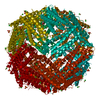
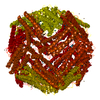
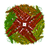
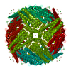
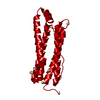
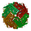
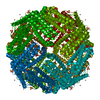
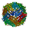
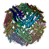
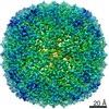
- Structure visualization
| Structure viewer | Molecule: MolmilJmol/JSmol |
|---|
- Downloads & links
-Download
| PDBx/mmCIF format | 1dat.cif.gz | 50.7 KB | Display | PDBx/mmCIF format |
|---|---|---|---|---|
| PDB format | pdb1dat.ent.gz | 37.8 KB | Display | PDB format |
| PDBx/mmJSON format | 1dat.json.gz | Tree view | PDBx/mmJSON format | |
| Others |  Other downloads Other downloads |
-Validation report
| Arichive directory | https://data.pdbj.org/pub/pdb/validation_reports/da/1datftp://data.pdbj.org/pub/pdb/validation_reports/da/1dat | HTTPS FTP |
|---|
-Related structure data
| Related structure data | 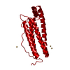 1ierS S: Starting model for refinement |
|---|---|
| Similar structure data |


-Links
 PDBj
PDBj


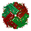
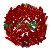
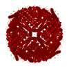
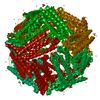
- Assembly
| Deposited unit | 
| ||||||||||||
|---|---|---|---|---|---|---|---|---|---|---|---|---|---|
| 1 | x 24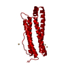
| ||||||||||||
| Unit cell |
| ||||||||||||
| Components on special symmetry positions |
|
-Components
| #1: Protein | Mass: 19856.385 Da / Num. of mol.: 1 Source method: isolated from a genetically manipulated source Source: (gene. exp.)  | ||
|---|---|---|---|
| #2: Chemical |   Mass: 112.411 Da / Num. of mol.: 3 / Source method: obtained synthetically / Formula: Cd Mass: 112.411 Da / Num. of mol.: 3 / Source method: obtained synthetically / Formula: Cd#3: Water | ChemComp-HOH / |  Mass: 18.015 Da / Num. of mol.: 106 / Source method: isolated from a natural source / Formula: H2O Mass: 18.015 Da / Num. of mol.: 106 / Source method: isolated from a natural source / Formula: H2O |
-Experimental details
-Experiment
| Experiment | Method: X-RAY DIFFRACTION / Number of used crystals: 1 |
|---|
- Sample preparation
| Crystal | Density Matthews: 3.26 Å3/Da / Density % sol: 62.2 % | ||||||||||||||||||||||||||||||
|---|---|---|---|---|---|---|---|---|---|---|---|---|---|---|---|---|---|---|---|---|---|---|---|---|---|---|---|---|---|---|---|
| Crystal grow | *PLUS Method: vapor diffusion, hanging drop | ||||||||||||||||||||||||||||||
| Components of the solutions | *PLUS
|
-Data collection
| Diffraction | Mean temperature: 293 K |
|---|---|
| Diffraction source | Source: SYNCHROTRON / Site: LURE  / Beamline: D41A / Wavelength: 1.375 / Beamline: D41A / Wavelength: 1.375 |
| Detector | Type: MARRESEARCH / Detector: IMAGE PLATE / Date: Jun 2, 1995 |
| Radiation | Monochromatic (M) / Laue (L): M / Scattering type: x-ray |
| Radiation wavelength | Wavelength: 1.375 Å / Relative weight: 1 |
| Reflection | Resolution: 2.05→16.05 Å / Num. obs: 16993 / % possible obs: 99.6 % / Observed criterion σ(I): 1 / Redundancy: 10.6 % / Biso Wilson estimate: 15.2 Å2 / Rsym value: 0.096 / Net I/σ(I): 6.4 |
| Reflection shell | Resolution: 2.05→2.17 Å / Redundancy: 9.3 % / Mean I/σ(I) obs: 2.7 / Rsym value: 0.278 / % possible all: 99.6 |
| Reflection | *PLUS Num. obs: 16931 / Num. measured all: 179720 / Rmerge(I) obs: 0.096 |
| Reflection shell | *PLUS % possible obs: 99.6 % / Rmerge(I) obs: 0.278 |
- Processing
| Software |
| ||||||||||||||||||||||||||||||||||||||||||||||||||||||||||||
|---|---|---|---|---|---|---|---|---|---|---|---|---|---|---|---|---|---|---|---|---|---|---|---|---|---|---|---|---|---|---|---|---|---|---|---|---|---|---|---|---|---|---|---|---|---|---|---|---|---|---|---|---|---|---|---|---|---|---|---|---|---|
| Refinement | Starting model: PDB ENTRY 1IER Resolution: 2.05→8 Å / σ(F): 4
| ||||||||||||||||||||||||||||||||||||||||||||||||||||||||||||
| Displacement parameters | Biso mean: 19.5 Å2 | ||||||||||||||||||||||||||||||||||||||||||||||||||||||||||||
| Refine analyze | Luzzati coordinate error obs: 0.2 Å | ||||||||||||||||||||||||||||||||||||||||||||||||||||||||||||
| Refinement step | Cycle: LAST / Resolution: 2.05→8 Å
| ||||||||||||||||||||||||||||||||||||||||||||||||||||||||||||
| Refine LS restraints |
| ||||||||||||||||||||||||||||||||||||||||||||||||||||||||||||
| LS refinement shell | Resolution: 2.05→2.15 Å
| ||||||||||||||||||||||||||||||||||||||||||||||||||||||||||||
| Software | *PLUS Name: X-PLOR / Version: 2.1 / Classification: refinement | ||||||||||||||||||||||||||||||||||||||||||||||||||||||||||||
| Refinement | *PLUS Rfactor obs: 0.179 / Rfactor Rwork: 0.179 | ||||||||||||||||||||||||||||||||||||||||||||||||||||||||||||
| Solvent computation | *PLUS | ||||||||||||||||||||||||||||||||||||||||||||||||||||||||||||
| Displacement parameters | *PLUS | ||||||||||||||||||||||||||||||||||||||||||||||||||||||||||||
| Refine LS restraints | *PLUS
|
