 Movie
Movie Controller

[English] 日本語
 Yorodumi
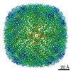
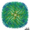
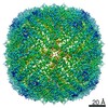
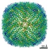
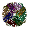
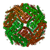
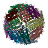
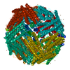
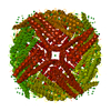
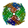
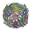
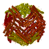
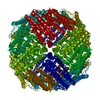
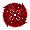
- PDB-7a6b: 1.33 A structure of human apoferritin obtained from Titan Mono- B... -
+ Open data

- Basic information
| Entry | Database: PDB / ID: 7a6b | |||||||||||||||||||||||||||
|---|---|---|---|---|---|---|---|---|---|---|---|---|---|---|---|---|---|---|---|---|---|---|---|---|---|---|---|---|
| Title | 1.33 A structure of human apoferritin obtained from Titan Mono- BCOR microscope | |||||||||||||||||||||||||||
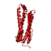
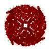
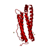
 Components Components | Ferritin heavy chain | |||||||||||||||||||||||||||
 Keywords Keywords | METAL BINDING PROTEIN / Apoferritin | |||||||||||||||||||||||||||
| Function / homology |  Function and homology information Function and homology informationiron ion sequestering activity / ferritin complex / Scavenging by Class A Receptors / Golgi Associated Vesicle Biogenesis / ferroxidase / negative regulation of ferroptosis / autolysosome / ferroxidase activity / negative regulation of fibroblast proliferation / ferric iron binding ...iron ion sequestering activity / ferritin complex / Scavenging by Class A Receptors / Golgi Associated Vesicle Biogenesis / ferroxidase / negative regulation of ferroptosis / autolysosome / ferroxidase activity / negative regulation of fibroblast proliferation / ferric iron binding / autophagosome / iron ion transport / ferrous iron binding / Iron uptake and transport / tertiary granule lumen / ficolin-1-rich granule lumen / intracellular iron ion homeostasis / immune response / iron ion binding / negative regulation of cell population proliferation / Neutrophil degranulation / extracellular exosome / extracellular region / identical protein binding / nucleus / cytoplasm / cytosol Similarity search - Function | |||||||||||||||||||||||||||
| Biological species |  Homo sapiens (human) Homo sapiens (human) | |||||||||||||||||||||||||||
| Method | ELECTRON MICROSCOPY / single particle reconstruction / cryo EM / Resolution: 1.33 Å | |||||||||||||||||||||||||||
 Authors Authors | Yip, K.M. / Fischer, N. / Chari, A. / Stark, H. | |||||||||||||||||||||||||||
| Funding support |  Germany, 2items Germany, 2items
| |||||||||||||||||||||||||||
 Citation Citation | Journal: Nature / Year: 2020 Title: Atomic-resolution protein structure determination by cryo-EM. Authors: Ka Man Yip / Niels Fischer / Elham Paknia / Ashwin Chari / Holger Stark / Abstract: Single-particle electron cryo-microscopy (cryo-EM) is a powerful method for solving the three-dimensional structures of biological macromolecules. The technological development of transmission ...Single-particle electron cryo-microscopy (cryo-EM) is a powerful method for solving the three-dimensional structures of biological macromolecules. The technological development of transmission electron microscopes, detectors and automated procedures in combination with user-friendly image processing software and ever-increasing computational power have made cryo-EM a successful and expanding technology over the past decade. At resolutions better than 4 Å, atomic model building starts to become possible, but the direct visualization of true atomic positions in protein structure determination requires much higher (better than 1.5 Å) resolution, which so far has not been attained by cryo-EM. The direct visualization of atom positions is essential for understanding the mechanisms of protein-catalysed chemical reactions, and for studying how drugs bind to and interfere with the function of proteins. Here we report a 1.25 Å-resolution structure of apoferritin obtained by cryo-EM with a newly developed electron microscope that provides, to our knowledge, unprecedented structural detail. Our apoferritin structure has almost twice the 3D information content of the current world record reconstruction (at 1.54 Å resolution). We can visualize individual atoms in a protein, see density for hydrogen atoms and image single-atom chemical modifications. Beyond the nominal improvement in resolution, we also achieve a substantial improvement in the quality of the cryo-EM density map, which is highly relevant for using cryo-EM in structure-based drug design. | |||||||||||||||||||||||||||
| History |
|
- Structure visualization
| Movie |
Movie viewer |
|---|---|
| Structure viewer | Molecule: MolmilJmol/JSmol |
- Downloads & links
-Download
| PDBx/mmCIF format | 7a6b.cif.gz | 1.9 MB | Display | PDBx/mmCIF format |
|---|---|---|---|---|
| PDB format | pdb7a6b.ent.gz | 1.6 MB | Display | PDB format |
| PDBx/mmJSON format | 7a6b.json.gz | Tree view | PDBx/mmJSON format | |
| Others |  Other downloads Other downloads |
-Validation report
| Arichive directory | https://data.pdbj.org/pub/pdb/validation_reports/a6/7a6bftp://data.pdbj.org/pub/pdb/validation_reports/a6/7a6b | HTTPS FTP |
|---|
-Related structure data
| Related structure data | 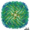 11669MC  6z6uC  6z9eC  6z9fC  7a6aC M: map data used to model this data C: citing same article ( |
|---|---|
| Similar structure data | |
| EM raw data | EMPIAR-10591 (Title: Atomic resolution structure of apoferritin from Titan Mono/BCorr microscope Data size: 41.7 TB Data #1: Single particle cryo-EM dataset of apoferritin from Titan Mono-BCorr microscope at 1.25 angstrom resolution [micrographs - multiframe]) |


-Links
 PDBj
PDBj


- Assembly
| Deposited unit | 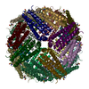
|
|---|---|
| 1 |
|
-Components
| #1: Protein | Mass: 21270.605 Da / Num. of mol.: 24 Source method: isolated from a genetically manipulated source Source: (gene. exp.) Homo sapiens (human) / Gene: FTH1, FTH, FTHL6, OK/SW-cl.84, PIG15 / Production host:  #2: Chemical | ChemComp-NA /   Mass: 22.990 Da / Num. of mol.: 32 / Source method: obtained synthetically / Formula: Na Mass: 22.990 Da / Num. of mol.: 32 / Source method: obtained synthetically / Formula: Na#3: Water | ChemComp-HOH / |  Mass: 18.015 Da / Num. of mol.: 3854 / Source method: isolated from a natural source / Formula: H2O Mass: 18.015 Da / Num. of mol.: 3854 / Source method: isolated from a natural source / Formula: H2OHas ligand of interest | N | Has protein modification | Y | |
|---|
-Experimental details
-Experiment
| Experiment | Method: ELECTRON MICROSCOPY |
|---|---|
| EM experiment | Aggregation state: PARTICLE / 3D reconstruction method: single particle reconstruction |
- Sample preparation
| Component | Name: Apoferritin / Type: ORGANELLE OR CELLULAR COMPONENT / Entity ID: #1 / Source: RECOMBINANT |
|---|---|
| Molecular weight | Experimental value: NO |
| Source (natural) | Organism: Homo sapiens (human) |
| Source (recombinant) | Organism: |
| Buffer solution | pH: 7.6 |
| Specimen | Conc.: 3.5 mg/ml / Embedding applied: NO / Shadowing applied: NO / Staining applied: NO / Vitrification applied: YES |
| Vitrification | Cryogen name: ETHANE |
- Electron microscopy imaging
| Experimental equipment |  Model: Titan Krios / Image courtesy: FEI Company |
|---|---|
| Microscopy | Model: FEI TITAN KRIOS |
| Electron gun | Electron source:  FIELD EMISSION GUN / Accelerating voltage: 300 kV / Illumination mode: FLOOD BEAM FIELD EMISSION GUN / Accelerating voltage: 300 kV / Illumination mode: FLOOD BEAM |
| Electron lens | Mode: BRIGHT FIELD |
| Image recording | Electron dose: 50 e/Å2 / Detector mode: COUNTING / Film or detector model: FEI FALCON III (4k x 4k) |
| EM imaging optics | Chromatic aberration corrector: TFS Monochromator / Spherical aberration corrector: CEOS B-COR |
- Processing
| EM software |
| |||||||||
|---|---|---|---|---|---|---|---|---|---|---|
| CTF correction | Type: PHASE FLIPPING ONLY | |||||||||
| 3D reconstruction | Resolution: 1.33 Å / Resolution method: FSC 0.143 CUT-OFF / Num. of particles: 1074000 / Symmetry type: POINT |
