Group: Data collection / Category: chem_comp_atom / chem_comp_bond
Remark 700
SHEET DETERMINATION METHOD: DSSP THE SHEETS PRESENTED AS "AA" IN EACH CHAIN ON SHEET RECORDS BELOW ... SHEET DETERMINATION METHOD: DSSP THE SHEETS PRESENTED AS "AA" IN EACH CHAIN ON SHEET RECORDS BELOW IS ACTUALLY AN 18-STRANDED BARREL THIS IS REPRESENTED BY A 19-STRANDED SHEET IN WHICH THE FIRST AND LAST STRANDS ARE IDENTICAL. THE SHEETS PRESENTED AS "BA" IN EACH CHAIN ON SHEET RECORDS BELOW IS ACTUALLY AN 17-STRANDED BARREL THIS IS REPRESENTED BY A 18-STRANDED SHEET IN WHICH THE FIRST AND LAST STRANDS ARE IDENTICAL. THE SHEETS PRESENTED AS "EA" IN EACH CHAIN ON SHEET RECORDS BELOW IS ACTUALLY AN 8-STRANDED BARREL THIS IS REPRESENTED BY A 9-STRANDED SHEET IN WHICH THE FIRST AND LAST STRANDS ARE IDENTICAL. THE SHEETS PRESENTED AS "FA" IN EACH CHAIN ON SHEET RECORDS BELOW IS ACTUALLY AN 8-STRANDED BARREL THIS IS REPRESENTED BY A 9-STRANDED SHEET IN WHICH THE FIRST AND LAST STRANDS ARE IDENTICAL. THE SHEETS PRESENTED AS "GA" IN EACH CHAIN ON SHEET RECORDS BELOW IS ACTUALLY AN 16-STRANDED BARREL THIS IS REPRESENTED BY A 17-STRANDED SHEET IN WHICH THE FIRST AND LAST STRANDS ARE IDENTICAL.
Mass: 18.015 Da / Num. of mol.: 159 / Source method: isolated from a natural source / Formula: H2O
Sequence details
THIS RECOMBINANT PLASMID CARRIES THE UGPG GENE PRECEDED BY A SEQUENCE CODING FOR SIX HISTIDINE ...THIS RECOMBINANT PLASMID CARRIES THE UGPG GENE PRECEDED BY A SEQUENCE CODING FOR SIX HISTIDINE RESIDUES FOLLOWED BY TWO RESIDUES, A GLYCINE AND A SERINE, BEFORE THE INITIATING METHIONINE.
-
Experimental details
-
Experiment
Experiment
Method: X-RAY DIFFRACTION / Number of used crystals: 1
-
Sample preparation
Crystal
Density Matthews: 2.55 Å3/Da / Density % sol: 51.7 %
Monochromator: SI(111) / Protocol: SINGLE WAVELENGTH / Monochromatic (M) / Laue (L): M / Scattering type: x-ray
Radiation wavelength
ID
Wavelength (Å)
Relative weight
1
0.97564
1
2
1.1398
1
Reflection
Resolution: 2.65→102 Å / Num. obs: 85566 / % possible obs: 100 % / Observed criterion σ(I): 2 / Redundancy: 5.7 % / Biso Wilson estimate: 50.3 Å2 / Rmerge(I) obs: 0.1 / Net I/σ(I): 14.1
Reflection shell
Resolution: 2.65→2.67 Å / Redundancy: 3.7 % / Rmerge(I) obs: 0.65 / Mean I/σ(I) obs: 2 / % possible all: 100
-
Processing
Software
Name
Version
Classification
CNS
1
refinement
MOSFLM
datareduction
SCALA
datascaling
SHARP
phasing
Refinement
Method to determine structure: MIRAS / Resolution: 2.65→74.9 Å / Rfactor Rfree error: 0.007 / Data cutoff high absF: 884457.43 / Isotropic thermal model: B-GROUP / Cross valid method: THROUGHOUT / σ(F): 0 Stereochemistry target values: MAXIMUM LIKELIHOOD TARGET USING AMPLITUDES Details: DIFFERENT NCS RESTRAINS USED FOR MAIN-CHAINS AND FOR SIDE-CHAINS. GROUP B-FACTOR REFINEMENT DOES NOT ALLOW NCS RESTRAINS. DISORDERED REGIONS WERE NOT INCLUDED IN THE REFINEMENT.
Rfactor
Num. reflection
% reflection
Selection details
Rfree
0.295
1992
2.6 %
SHELLS
Rwork
0.245
-
-
-
obs
0.245
76179
100 %
-
Solvent computation
Solvent model: CNS BULK SOLVENT MODEL USED / Bsol: 45.5985 Å2 / ksol: 0.351613 e/Å3
Displacement parameters
Biso mean: 54.23 Å2
Baniso -1
Baniso -2
Baniso -3
1-
5.01 Å2
0 Å2
-6.83 Å2
2-
-
-5.13 Å2
0 Å2
3-
-
-
0.11 Å2
Refine analyze
Free
Obs
Luzzati coordinate error
0.48 Å
0.38 Å
Luzzati sigma a
0.52 Å
0.43 Å
Refinement step
Cycle: LAST / Resolution: 2.65→74.9 Å
Protein
Nucleic acid
Ligand
Solvent
Total
Num. atoms
16661
0
128
159
16948
Refine LS restraints
Refine-ID
Type
Dev ideal
X-RAY DIFFRACTION
c_bond_d
0.012
X-RAY DIFFRACTION
c_bond_d_na
X-RAY DIFFRACTION
c_bond_d_prot
X-RAY DIFFRACTION
c_angle_d
X-RAY DIFFRACTION
c_angle_d_na
X-RAY DIFFRACTION
c_angle_d_prot
X-RAY DIFFRACTION
c_angle_deg
1.84
X-RAY DIFFRACTION
c_angle_deg_na
X-RAY DIFFRACTION
c_angle_deg_prot
X-RAY DIFFRACTION
c_dihedral_angle_d
X-RAY DIFFRACTION
c_dihedral_angle_d_na
X-RAY DIFFRACTION
c_dihedral_angle_d_prot
X-RAY DIFFRACTION
c_improper_angle_d
X-RAY DIFFRACTION
c_improper_angle_d_na
X-RAY DIFFRACTION
c_improper_angle_d_prot
X-RAY DIFFRACTION
c_mcbond_it
X-RAY DIFFRACTION
c_mcangle_it
X-RAY DIFFRACTION
c_scbond_it
X-RAY DIFFRACTION
c_scangle_it
Refine LS restraints NCS
NCS model details: RESTRAINS / Rms dev position: 2 Å / Weight position: 10
LS refinement shell
Resolution: 2.75→2.77 Å / Total num. of bins used: 39
Rfactor
Num. reflection
% reflection
Rfree
0.403
492
0.246 %
Rwork
0.346
1500
-
obs
-
-
100 %
Xplor file
Serial no: 1 / Param file: PROTEIN_REP.PARAM
+
About Yorodumi
-
News
-
Feb 9, 2022. New format data for meta-information of EMDB entries
New format data for meta-information of EMDB entries
Version 3 of the EMDB header file is now the official format.
The previous official version 1.9 will be removed from the archive.
In the structure databanks used in Yorodumi, some data are registered as the other names, "COVID-19 virus" and "2019-nCoV". Here are the details of the virus and the list of structure data.
Jan 31, 2019. EMDB accession codes are about to change! (news from PDBe EMDB page)
EMDB accession codes are about to change! (news from PDBe EMDB page)
The allocation of 4 digits for EMDB accession codes will soon come to an end. Whilst these codes will remain in use, new EMDB accession codes will include an additional digit and will expand incrementally as the available range of codes is exhausted. The current 4-digit format prefixed with “EMD-” (i.e. EMD-XXXX) will advance to a 5-digit format (i.e. EMD-XXXXX), and so on. It is currently estimated that the 4-digit codes will be depleted around Spring 2019, at which point the 5-digit format will come into force.
The EM Navigator/Yorodumi systems omit the EMD- prefix.
Related info.:Q: What is EMD? / ID/Accession-code notation in Yorodumi/EM Navigator
Yorodumi is a browser for structure data from EMDB, PDB, SASBDB, etc.
This page is also the successor to EM Navigator detail page, and also detail information page/front-end page for Omokage search.
The word "yorodu" (or yorozu) is an old Japanese word meaning "ten thousand". "mi" (miru) is to see.
Related info.:EMDB / PDB / SASBDB / Comparison of 3 databanks / Yorodumi Search / Aug 31, 2016. New EM Navigator & Yorodumi / Yorodumi Papers / Jmol/JSmol / Function and homology information / Changes in new EM Navigator and Yorodumi
 Movie
Movie Controller

 Yorodumi
 Open data
 Basic information
 Components
 Keywords
 Function and homology information
 SPHINGOMONAS ELODEA (bacteria)
 X-RAY DIFFRACTION /
  Authors
 Citation
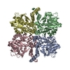
 Structure visualization
 Downloads & links
 Other downloads


 PDBj
PDBj

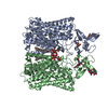
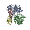
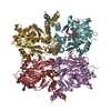
 Assembly
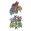

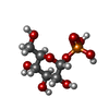
 Type: D-saccharide / Mass: 260.136 Da / Num. of mol.: 8
 Mass: 18.015 Da / Num. of mol.: 159 / Source method: isolated from a natural source / Formula: H2O
 Sample preparation
 / Beamline: ID29 / Wavelength: 0.97564, 1.13980
 Processing