 Movie
Movie Controller

[English] 日本語
 Yorodumi
- PDB-6mnu: Crystal structure of Yersinia pestis UDP-glucose pyrophosphorylase -
+ Open data

- Basic information
| Entry | Database: PDB / ID: 6mnu | ||||||
|---|---|---|---|---|---|---|---|
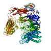
| Title | Crystal structure of Yersinia pestis UDP-glucose pyrophosphorylase | ||||||
 Components Components | UTP--glucose-1-phosphate uridylyltransferase | ||||||
 Keywords Keywords | TRANSFERASE / UDP-glucose pyrophosphorylase / plague / Yersinia pestis | ||||||
| Function / homology |  Function and homology information Function and homology informationUTP-glucose-1-phosphate uridylyltransferase / UTP:glucose-1-phosphate uridylyltransferase activity / UDP-alpha-D-glucose metabolic process / biosynthetic process Similarity search - Function | ||||||
| Biological species |   Yersinia pestis (bacteria) Yersinia pestis (bacteria) | ||||||
| Method |  X-RAY DIFFRACTION / SYNCHROTRON / MOLECULAR REPLACEMENT / Resolution: 2.172 Å X-RAY DIFFRACTION / SYNCHROTRON / MOLECULAR REPLACEMENT / Resolution: 2.172 Å | ||||||
 Authors Authors | Gibbs, M.E. / Lountos, G.T. / Gumpena, R. / Waugh, D.S. | ||||||
 Citation Citation | Journal: Acta Crystallogr.,Sect.F / Year: 2019 Title: Crystal structure of UDP-glucose pyrophosphorylase from Yersinia pestis, a potential therapeutic target against plague. Authors: Gibbs, M.E. / Lountos, G.T. / Gumpena, R. / Waugh, D.S. | ||||||
| History |
|
- Structure visualization
| Structure viewer | Molecule: MolmilJmol/JSmol |
|---|
- Downloads & links
-Download
| PDBx/mmCIF format | 6mnu.cif.gz | 236.4 KB | Display | PDBx/mmCIF format |
|---|---|---|---|---|
| PDB format | pdb6mnu.ent.gz | 189.6 KB | Display | PDB format |
| PDBx/mmJSON format | 6mnu.json.gz | Tree view | PDBx/mmJSON format | |
| Others |  Other downloads Other downloads |
-Validation report
| Arichive directory | https://data.pdbj.org/pub/pdb/validation_reports/mn/6mnuftp://data.pdbj.org/pub/pdb/validation_reports/mn/6mnu | HTTPS FTP |
|---|
-Related structure data
| Related structure data | 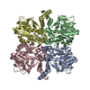 2e3dS S: Starting model for refinement |
|---|---|
| Similar structure data |


-Links
 PDBj
PDBj

- Assembly
| Deposited unit | 
| ||||||||
|---|---|---|---|---|---|---|---|---|---|
| 1 |
| ||||||||
| Unit cell |
|
-Components
| #1: Protein | Mass: 32308.621 Da / Num. of mol.: 4 Source method: isolated from a genetically manipulated source Source: (gene. exp.) Yersinia pestis (bacteria)Gene: galU, galU1, y2631, YP_1428, DBB30_10560, EGT45_14485, NCTC144_03080 Plasmid: pMG3038 / Production host: References: UniProt: A0A2S9PFH5, UniProt: A0A3N4B738*PLUS, UTP-glucose-1-phosphate uridylyltransferase #2: Chemical |   Mass: 62.068 Da / Num. of mol.: 2 / Source method: obtained synthetically / Formula: C2H6O2 Mass: 62.068 Da / Num. of mol.: 2 / Source method: obtained synthetically / Formula: C2H6O2#3: Water | ChemComp-HOH / |  Mass: 18.015 Da / Num. of mol.: 623 / Source method: isolated from a natural source / Formula: H2O Mass: 18.015 Da / Num. of mol.: 623 / Source method: isolated from a natural source / Formula: H2O |
|---|
-Experimental details
-Experiment
| Experiment | Method: X-RAY DIFFRACTION / Number of used crystals: 1 |
|---|
- Sample preparation
| Crystal | Density Matthews: 2.28 Å3/Da / Density % sol: 46.15 % |
|---|---|
| Crystal grow | Temperature: 292 K / Method: vapor diffusion, hanging drop / pH: 6 Details: 0.1M MES pH 6.0 20% PEG Low Smear (PEG 1000, PEG400, PEG600, PEG MME 500), 10 mM spermidine |
-Data collection
| Diffraction | Mean temperature: 100 K / Serial crystal experiment: N |
|---|---|
| Diffraction source | Source: SYNCHROTRON / Site: APS  / Beamline: 22-BM / Wavelength: 1 Å / Beamline: 22-BM / Wavelength: 1 Å |
| Detector | Type: RAYONIX MX300-HS / Detector: CCD / Date: Aug 5, 2017 |
| Radiation | Protocol: SINGLE WAVELENGTH / Monochromatic (M) / Laue (L): M / Scattering type: x-ray |
| Radiation wavelength | Wavelength: 1 Å / Relative weight: 1 |
| Reflection | Resolution: 2.17→50 Å / Num. obs: 61347 / % possible obs: 97.6 % / Redundancy: 4.6 % / Rpim(I) all: 0.041 / Rrim(I) all: 0.09 / Rsym value: 0.08 / Net I/σ(I): 17.1 |
| Reflection shell | Resolution: 2.17→2.21 Å / Redundancy: 4.2 % / Mean I/σ(I) obs: 2.1 / Num. unique obs: 3054 / CC1/2: 0.774 / Rpim(I) all: 0.343 / Rrim(I) all: 0.722 / Rsym value: 0.631 / % possible all: 99.1 |
- Processing
| Software |
| |||||||||||||||||||||||||||||||||||||||||||||||||||||||||||||||||||||||||||||||||||||||||||||||||||||||||
|---|---|---|---|---|---|---|---|---|---|---|---|---|---|---|---|---|---|---|---|---|---|---|---|---|---|---|---|---|---|---|---|---|---|---|---|---|---|---|---|---|---|---|---|---|---|---|---|---|---|---|---|---|---|---|---|---|---|---|---|---|---|---|---|---|---|---|---|---|---|---|---|---|---|---|---|---|---|---|---|---|---|---|---|---|---|---|---|---|---|---|---|---|---|---|---|---|---|---|---|---|---|---|---|---|---|---|
| Refinement | Method to determine structure: MOLECULAR REPLACEMENT Starting model: 2E3D Resolution: 2.172→26.709 Å / SU ML: 0.26 / Cross valid method: FREE R-VALUE / σ(F): 1.34 / Phase error: 23.38
| |||||||||||||||||||||||||||||||||||||||||||||||||||||||||||||||||||||||||||||||||||||||||||||||||||||||||
| Solvent computation | Shrinkage radii: 0.9 Å / VDW probe radii: 1.11 Å | |||||||||||||||||||||||||||||||||||||||||||||||||||||||||||||||||||||||||||||||||||||||||||||||||||||||||
| Refinement step | Cycle: LAST / Resolution: 2.172→26.709 Å
| |||||||||||||||||||||||||||||||||||||||||||||||||||||||||||||||||||||||||||||||||||||||||||||||||||||||||
| Refine LS restraints |
| |||||||||||||||||||||||||||||||||||||||||||||||||||||||||||||||||||||||||||||||||||||||||||||||||||||||||
| LS refinement shell |
|
