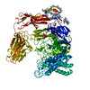
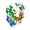
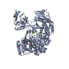
Mass: 109100.414 Da / Num. of mol.: 1 Source method: isolated from a genetically manipulated source Details: The last domain is very poorly defined, some fragments are described as UNK residues Source: (gene. exp.) Paenibacillus sp. (bacteria) / Production host: Bacillus subtilis (bacteria) / References: UniProt: A0A3F2YM17*PLUS
Protocol: SINGLE WAVELENGTH / Monochromatic (M) / Laue (L): M / Scattering type: x-ray
Radiation wavelength
Wavelength: 1.07 Å / Relative weight: 1
Reflection
Resolution: 2→52.29 Å / Num. obs: 88513 / % possible obs: 100 % / Redundancy: 9.9 % / CC1/2: 0.999 / Rmerge(I) obs: 0.082 / Net I/σ(I): 16
Reflection shell
Resolution: 2→2.05 Å / Rmerge(I) obs: 0.64 / Mean I/σ(I) obs: 3.5 / CC1/2: 0.876 / % possible all: 100
-
Processing
Software
Name
Version
Classification
REFMAC
5.8.0189
refinement
xia2
datareduction
Aimless
datascaling
CRANK2
phasing
Refinement
Method to determine structure: SAD / Resolution: 2→52.29 Å / Cor.coef. Fo:Fc: 0.96 / Cor.coef. Fo:Fc free: 0.945 / SU B: 6.073 / SU ML: 0.094 / Cross valid method: THROUGHOUT / ESU R: 0.143 / ESU R Free: 0.135 / Details: HYDROGENS HAVE BEEN ADDED IN THE RIDING POSITIONS
Rfactor
Num. reflection
% reflection
Selection details
Rfree
0.20976
4435
5 %
RANDOM
Rwork
0.17272
-
-
-
obs
0.17458
84008
99.98 %
-
Solvent computation
Ion probe radii: 0.8 Å / Shrinkage radii: 0.8 Å / VDW probe radii: 1.2 Å
 Movie
Movie Controller

 Yorodumi
 Open data
 Basic information
 Components
 Keywords
 Function and homology information
 Paenibacillus sp. (bacteria)
 X-RAY DIFFRACTION /
  Authors
 Citation
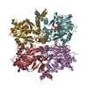
 Structure visualization
 Downloads & links
 Other downloads


 PDBj
PDBj


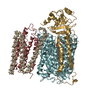
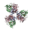
 Assembly


 Mass: 195.078 Da / Num. of mol.: 2 / Source method: obtained synthetically / Formula: Pt
 Mass: 282.334 Da / Num. of mol.: 1 / Source method: obtained synthetically / Formula: C11H26N2O6 / Comment: pH buffer*YM
 Mass: 58.082 Da / Num. of mol.: 2 / Source method: obtained synthetically / Formula: CNS
 Mass: 40.078 Da / Num. of mol.: 5 / Source method: obtained synthetically / Formula: Ca
 Mass: 22.990 Da / Num. of mol.: 3 / Source method: obtained synthetically / Formula: Na
 Mass: 35.453 Da / Num. of mol.: 1 / Source method: obtained synthetically / Formula: Cl
 Mass: 102.046 Da / Num. of mol.: 1 / Source method: obtained synthetically / Formula: C3H2O4
 Sample preparation
 / Beamline: I03 / Wavelength: 1.07 Å
 Processing