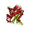
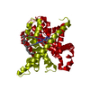
 Movie
Movie Controller


+ Open data

- Basic information
| Entry | Database: PDB / ID: 6ikx | ||||||
|---|---|---|---|---|---|---|---|
| Title | UDP-glucose pyrophosphorylase from acinetobacter baumanii | ||||||
 Components Components | UTP--glucose-1-phosphate uridylyltransferase | ||||||
 Keywords Keywords | TRANSFERASE / UDP-glucose pyrophosphorylase from acinetobacter baumanii | ||||||
| Function / homology |  Function and homology information Function and homology informationUTP-glucose-1-phosphate uridylyltransferase / UTP:glucose-1-phosphate uridylyltransferase activity / UDP-alpha-D-glucose metabolic process Similarity search - Function | ||||||
| Biological species |  Acinetobacter baumannii (bacteria) Acinetobacter baumannii (bacteria) | ||||||
| Method |  X-RAY DIFFRACTION / SYNCHROTRON / MOLECULAR REPLACEMENT / Resolution: 2.2 Å X-RAY DIFFRACTION / SYNCHROTRON / MOLECULAR REPLACEMENT / Resolution: 2.2 Å | ||||||
 Authors Authors | Lee, J.H. / Kang, L.W. | ||||||
 Citation Citation | Journal: To be published Title: UDP-glucose pyrophosphorylase from acinetobacter baumanii Authors: Kang, L.W. | ||||||
| History |
|
- Structure visualization
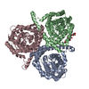
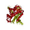
| Structure viewer | Molecule: MolmilJmol/JSmol |
|---|
- Downloads & links
-Download
| PDBx/mmCIF format | 6ikx.cif.gz | 124.5 KB | Display | PDBx/mmCIF format |
|---|---|---|---|---|
| PDB format | pdb6ikx.ent.gz | 95.8 KB | Display | PDB format |
| PDBx/mmJSON format | 6ikx.json.gz | Tree view | PDBx/mmJSON format | |
| Others |  Other downloads Other downloads |
-Validation report
| Arichive directory | https://data.pdbj.org/pub/pdb/validation_reports/ik/6ikxftp://data.pdbj.org/pub/pdb/validation_reports/ik/6ikx | HTTPS FTP |
|---|
-Related structure data
| Related structure data | 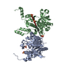 5j49S S: Starting model for refinement |
|---|---|
| Similar structure data |


-Links
 PDBj
PDBj

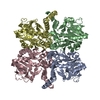
- Assembly
| Deposited unit | 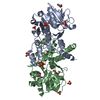
| ||||||||
|---|---|---|---|---|---|---|---|---|---|
| 1 | 
| ||||||||
| Unit cell |
|
-Components
| #1: Protein | Mass: 31665.459 Da / Num. of mol.: 2 / Mutation: Q286L Source method: isolated from a genetically manipulated source Source: (gene. exp.) Acinetobacter baumannii (bacteria)Gene: galU, B9X91_19205, CBI29_00108, CSB70_3798, DVA79_14980 Plasmid: pET11a / Production host: References: UniProt: X2KZJ9, UTP-glucose-1-phosphate uridylyltransferase #2: Chemical |   Mass: 92.094 Da / Num. of mol.: 2 / Source method: obtained synthetically / Formula: C3H8O3 Mass: 92.094 Da / Num. of mol.: 2 / Source method: obtained synthetically / Formula: C3H8O3#3: Chemical | ChemComp-SO4 /   Mass: 96.063 Da / Num. of mol.: 12 / Source method: obtained synthetically / Formula: SO4 Mass: 96.063 Da / Num. of mol.: 12 / Source method: obtained synthetically / Formula: SO4#4: Chemical |   Mass: 62.068 Da / Num. of mol.: 3 / Source method: obtained synthetically / Formula: C2H6O2 Mass: 62.068 Da / Num. of mol.: 3 / Source method: obtained synthetically / Formula: C2H6O2#5: Water | ChemComp-HOH / |  Mass: 18.015 Da / Num. of mol.: 75 / Source method: isolated from a natural source / Formula: H2O Mass: 18.015 Da / Num. of mol.: 75 / Source method: isolated from a natural source / Formula: H2O |
|---|
-Experimental details
-Experiment
| Experiment | Method: X-RAY DIFFRACTION / Number of used crystals: 1 |
|---|
- Sample preparation
| Crystal | Density Matthews: 3.06 Å3/Da / Density % sol: 59.82 % / Mosaicity: 0.407 ° |
|---|---|
| Crystal grow | Temperature: 287 K / Method: evaporation / pH: 7.5 Details: 1.5M Ammonium citrate, 0.1M BIS-TRIS pH 6.13 and 0.1M NaCl |
-Data collection
| Diffraction | Mean temperature: 100 K / Serial crystal experiment: N | |||||||||||||||||||||||||||||||||||||||||||||||||||||||||||||||||||||||||||||||||||||||||||||||||||||||||||||||||||||||||||||||||||||||||||||||||||||||||||||||||||||||||||||||||||||||||||||
|---|---|---|---|---|---|---|---|---|---|---|---|---|---|---|---|---|---|---|---|---|---|---|---|---|---|---|---|---|---|---|---|---|---|---|---|---|---|---|---|---|---|---|---|---|---|---|---|---|---|---|---|---|---|---|---|---|---|---|---|---|---|---|---|---|---|---|---|---|---|---|---|---|---|---|---|---|---|---|---|---|---|---|---|---|---|---|---|---|---|---|---|---|---|---|---|---|---|---|---|---|---|---|---|---|---|---|---|---|---|---|---|---|---|---|---|---|---|---|---|---|---|---|---|---|---|---|---|---|---|---|---|---|---|---|---|---|---|---|---|---|---|---|---|---|---|---|---|---|---|---|---|---|---|---|---|---|---|---|---|---|---|---|---|---|---|---|---|---|---|---|---|---|---|---|---|---|---|---|---|---|---|---|---|---|---|---|---|---|---|---|
| Diffraction source | Source: SYNCHROTRON / Site: PAL/PLS  / Beamline: 5C (4A) / Wavelength: 0.9794 Å / Beamline: 5C (4A) / Wavelength: 0.9794 Å | |||||||||||||||||||||||||||||||||||||||||||||||||||||||||||||||||||||||||||||||||||||||||||||||||||||||||||||||||||||||||||||||||||||||||||||||||||||||||||||||||||||||||||||||||||||||||||||
| Detector | Type: ADSC QUANTUM 315r / Detector: CCD / Date: May 2, 2018 | |||||||||||||||||||||||||||||||||||||||||||||||||||||||||||||||||||||||||||||||||||||||||||||||||||||||||||||||||||||||||||||||||||||||||||||||||||||||||||||||||||||||||||||||||||||||||||||
| Radiation | Protocol: SINGLE WAVELENGTH / Monochromatic (M) / Laue (L): M / Scattering type: x-ray | |||||||||||||||||||||||||||||||||||||||||||||||||||||||||||||||||||||||||||||||||||||||||||||||||||||||||||||||||||||||||||||||||||||||||||||||||||||||||||||||||||||||||||||||||||||||||||||
| Radiation wavelength | Wavelength: 0.9794 Å / Relative weight: 1 | |||||||||||||||||||||||||||||||||||||||||||||||||||||||||||||||||||||||||||||||||||||||||||||||||||||||||||||||||||||||||||||||||||||||||||||||||||||||||||||||||||||||||||||||||||||||||||||
| Reflection | Resolution: 2.2→50 Å / Num. obs: 39722 / % possible obs: 99.4 % / Redundancy: 11.2 % / Rmerge(I) obs: 0.107 / Rpim(I) all: 0.029 / Rrim(I) all: 0.112 / Χ2: 2.492 / Net I/σ(I): 10 / Num. measured all: 446063 | |||||||||||||||||||||||||||||||||||||||||||||||||||||||||||||||||||||||||||||||||||||||||||||||||||||||||||||||||||||||||||||||||||||||||||||||||||||||||||||||||||||||||||||||||||||||||||||
| Reflection shell | Diffraction-ID: 1
|
- Processing
| Software |
| ||||||||||||||||||||||||||||||||||||||||||||||||||||||||||||
|---|---|---|---|---|---|---|---|---|---|---|---|---|---|---|---|---|---|---|---|---|---|---|---|---|---|---|---|---|---|---|---|---|---|---|---|---|---|---|---|---|---|---|---|---|---|---|---|---|---|---|---|---|---|---|---|---|---|---|---|---|---|
| Refinement | Method to determine structure: MOLECULAR REPLACEMENT Starting model: 5J49 Resolution: 2.2→47.72 Å / Cor.coef. Fo:Fc: 0.959 / Cor.coef. Fo:Fc free: 0.909 / SU B: 8.956 / SU ML: 0.206 / Cross valid method: THROUGHOUT / σ(F): 0 / ESU R: 0.229 / ESU R Free: 0.227 / Stereochemistry target values: MAXIMUM LIKELIHOOD Details: HYDROGENS HAVE BEEN ADDED IN THE RIDING POSITIONS U VALUES : REFINED INDIVIDUALLY
| ||||||||||||||||||||||||||||||||||||||||||||||||||||||||||||
| Solvent computation | Ion probe radii: 0.8 Å / Shrinkage radii: 0.8 Å / VDW probe radii: 1.2 Å / Solvent model: MASK | ||||||||||||||||||||||||||||||||||||||||||||||||||||||||||||
| Displacement parameters | Biso max: 185.02 Å2 / Biso mean: 51.471 Å2 / Biso min: 30.57 Å2
| ||||||||||||||||||||||||||||||||||||||||||||||||||||||||||||
| Refinement step | Cycle: final / Resolution: 2.2→47.72 Å
| ||||||||||||||||||||||||||||||||||||||||||||||||||||||||||||
| Refine LS restraints |
| ||||||||||||||||||||||||||||||||||||||||||||||||||||||||||||
| LS refinement shell | Resolution: 2.201→2.258 Å / Rfactor Rfree error: 0 / Total num. of bins used: 20
|
