 Movie
Movie Controller


+ Open data

- Basic information
| Entry | Database: PDB / ID: 5u9w | ||||||
|---|---|---|---|---|---|---|---|
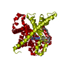
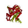
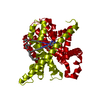
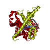
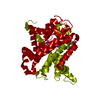
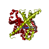
| Title | Structure of CNTnw N149L in the intermediate 3 state | ||||||
 Components Components | Nucleoside permease | ||||||
 Keywords Keywords | TRANSPORT PROTEIN / transporter / nucleoside / elevator-type alternate access | ||||||
| Function / homology |  Function and homology information Function and homology informationnucleoside transmembrane transporter activity / symporter activity / plasma membrane Similarity search - Function | ||||||
| Biological species |  Neisseria wadsworthii 9715 (bacteria) Neisseria wadsworthii 9715 (bacteria) | ||||||
| Method |  X-RAY DIFFRACTION / SYNCHROTRON / MOLECULAR REPLACEMENT / Resolution: 3.555 Å X-RAY DIFFRACTION / SYNCHROTRON / MOLECULAR REPLACEMENT / Resolution: 3.555 Å | ||||||
 Authors Authors | Hirschi, M. / Johnson, Z.L. / Lee, S.-Y. | ||||||
 Citation Citation | Journal: Nature / Year: 2017 Title: Visualizing multistep elevator-like transitions of a nucleoside transporter. Authors: Hirschi, M. / Johnson, Z.L. / Lee, S.Y. | ||||||
| History |
|
- Structure visualization
| Structure viewer | Molecule: MolmilJmol/JSmol |
|---|
- Downloads & links
-Download
| PDBx/mmCIF format | 5u9w.cif.gz | 229 KB | Display | PDBx/mmCIF format |
|---|---|---|---|---|
| PDB format | pdb5u9w.ent.gz | 180.7 KB | Display | PDB format |
| PDBx/mmJSON format | 5u9w.json.gz | Tree view | PDBx/mmJSON format | |
| Others |  Other downloads Other downloads |
-Validation report
| Arichive directory | https://data.pdbj.org/pub/pdb/validation_reports/u9/5u9wftp://data.pdbj.org/pub/pdb/validation_reports/u9/5u9w | HTTPS FTP |
|---|
-Related structure data
| Related structure data |  5l24C  5l26SC 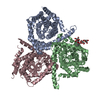 5l27C  5l2aC  5l2bC S: Starting model for refinement C: citing same article ( |
|---|---|
| Similar structure data |


-Links
 PDBj
PDBj- Assembly
| Deposited unit | 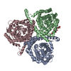
| ||||||||
|---|---|---|---|---|---|---|---|---|---|
| 1 |
| ||||||||
| Unit cell |
|
-Components
| #1: Protein | Mass: 44673.602 Da / Num. of mol.: 3 / Mutation: N149L Source method: isolated from a genetically manipulated source Source: (gene. exp.) Neisseria wadsworthii 9715 (bacteria) / Gene: nupC, HMPREF9370_1765 / Production host: #2: Chemical |   Mass: 949.082 Da / Num. of mol.: 2 / Source method: obtained synthetically / Formula: C43H80O22 Mass: 949.082 Da / Num. of mol.: 2 / Source method: obtained synthetically / Formula: C43H80O22 |
|---|
-Experimental details
-Experiment
| Experiment | Method: X-RAY DIFFRACTION / Number of used crystals: 1 |
|---|
- Sample preparation
| Crystal | Density Matthews: 3.66 Å3/Da / Density % sol: 66.4 % |
|---|---|
| Crystal grow | Temperature: 290 K / Method: vapor diffusion, sitting drop / pH: 7 / Details: 10mM Mg(OAc)2, 25% PEG400 |
-Data collection
| Diffraction | Mean temperature: 100 K |
|---|---|
| Diffraction source | Source: SYNCHROTRON / Site: APS  / Beamline: 22-ID / Wavelength: 0.97779 Å / Beamline: 22-ID / Wavelength: 0.97779 Å |
| Detector | Type: MARMOSAIC 300 mm CCD / Detector: CCD / Date: Oct 8, 2016 |
| Radiation | Protocol: SINGLE WAVELENGTH / Monochromatic (M) / Laue (L): M / Scattering type: x-ray |
| Radiation wavelength | Wavelength: 0.97779 Å / Relative weight: 1 |
| Reflection | Resolution: 3.55→30 Å / Num. obs: 24655 / % possible obs: 99.9 % / Redundancy: 8.4 % / CC1/2: 0.93 / Rpim(I) all: 0.05 / Net I/σ(I): 16.4 |
| Reflection shell | Resolution: 3.55→3.61 Å / Redundancy: 8.5 % / Mean I/σ(I) obs: 1.7 / CC1/2: 0.56 / Rpim(I) all: 0.69 / % possible all: 100 |
- Processing
| Software |
| ||||||||||||||||||||||||||||||||||||||||||||||||||||||||||||||||||||||||||||||||||||||||||||||||||||||||||||||||||||||||||||||||||||||||||||||||||||||||||||||||||||||||||||||||||||||
|---|---|---|---|---|---|---|---|---|---|---|---|---|---|---|---|---|---|---|---|---|---|---|---|---|---|---|---|---|---|---|---|---|---|---|---|---|---|---|---|---|---|---|---|---|---|---|---|---|---|---|---|---|---|---|---|---|---|---|---|---|---|---|---|---|---|---|---|---|---|---|---|---|---|---|---|---|---|---|---|---|---|---|---|---|---|---|---|---|---|---|---|---|---|---|---|---|---|---|---|---|---|---|---|---|---|---|---|---|---|---|---|---|---|---|---|---|---|---|---|---|---|---|---|---|---|---|---|---|---|---|---|---|---|---|---|---|---|---|---|---|---|---|---|---|---|---|---|---|---|---|---|---|---|---|---|---|---|---|---|---|---|---|---|---|---|---|---|---|---|---|---|---|---|---|---|---|---|---|---|---|---|---|---|
| Refinement | Method to determine structure: MOLECULAR REPLACEMENT Starting model: 5L26 Resolution: 3.555→29.964 Å / SU ML: 0.54 / Cross valid method: FREE R-VALUE / σ(F): 1.35 / Phase error: 29.8
| ||||||||||||||||||||||||||||||||||||||||||||||||||||||||||||||||||||||||||||||||||||||||||||||||||||||||||||||||||||||||||||||||||||||||||||||||||||||||||||||||||||||||||||||||||||||
| Solvent computation | Shrinkage radii: 0.9 Å / VDW probe radii: 1.11 Å | ||||||||||||||||||||||||||||||||||||||||||||||||||||||||||||||||||||||||||||||||||||||||||||||||||||||||||||||||||||||||||||||||||||||||||||||||||||||||||||||||||||||||||||||||||||||
| Refinement step | Cycle: LAST / Resolution: 3.555→29.964 Å
| ||||||||||||||||||||||||||||||||||||||||||||||||||||||||||||||||||||||||||||||||||||||||||||||||||||||||||||||||||||||||||||||||||||||||||||||||||||||||||||||||||||||||||||||||||||||
| Refine LS restraints |
| ||||||||||||||||||||||||||||||||||||||||||||||||||||||||||||||||||||||||||||||||||||||||||||||||||||||||||||||||||||||||||||||||||||||||||||||||||||||||||||||||||||||||||||||||||||||
| LS refinement shell |
|
