 Movie
Movie Controller

[English] 日本語
 Yorodumi
- PDB-2rh7: Crystal Structures of the Luciferase and Green Fluorescent Protei... -
+ Open data

- Basic information
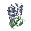
| Entry | Database: PDB / ID: 2rh7 | ||||||||||||
|---|---|---|---|---|---|---|---|---|---|---|---|---|---|
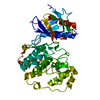
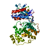
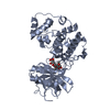
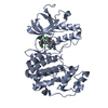
| Title | Crystal Structures of the Luciferase and Green Fluorescent Protein from Renilla Reniformis | ||||||||||||
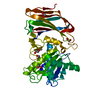
 Components Components | Green fluorescent protein | ||||||||||||
 Keywords Keywords | FLUORESCENT PROTEIN | ||||||||||||
| Function / homology |  Function and homology information Function and homology information | ||||||||||||
| Biological species |  Renilla reniformis (sea pansy) Renilla reniformis (sea pansy) | ||||||||||||
| Method |  X-RAY DIFFRACTION / SYNCHROTRON / MOLECULAR REPLACEMENT / Resolution: 1.5 Å X-RAY DIFFRACTION / SYNCHROTRON / MOLECULAR REPLACEMENT / Resolution: 1.5 Å | ||||||||||||
 Authors Authors | Loening, A.M. / Fenn, T.D. / Gambhir, S.S. | ||||||||||||
 Citation Citation | Journal: J.Mol.Biol. / Year: 2007 Title: Crystal Structures of the Luciferase and Green Fluorescent Protein from Renilla reniformis. Authors: Loening, A.M. / Fenn, T.D. / Gambhir, S.S. | ||||||||||||
| History |
|
- Structure visualization
| Structure viewer | Molecule: MolmilJmol/JSmol |
|---|
- Downloads & links
-Download
| PDBx/mmCIF format | 2rh7.cif.gz | 347 KB | Display | PDBx/mmCIF format |
|---|---|---|---|---|
| PDB format | pdb2rh7.ent.gz | 285.9 KB | Display | PDB format |
| PDBx/mmJSON format | 2rh7.json.gz | Tree view | PDBx/mmJSON format | |
| Others |  Other downloads Other downloads |
-Validation report
| Arichive directory | https://data.pdbj.org/pub/pdb/validation_reports/rh/2rh7ftp://data.pdbj.org/pub/pdb/validation_reports/rh/2rh7 | HTTPS FTP |
|---|
-Related structure data
| Related structure data | 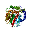 2psdC 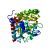 2pseC 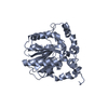 2psfC 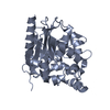 2pshC 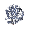 2psjC 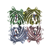 1ggxS  1mouS  1movS 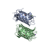 1xssS S: Starting model for refinement C: citing same article ( |
|---|---|
| Similar structure data |


-Links
 PDBj
PDBj

- Assembly
| Deposited unit | 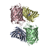
| ||||||||
|---|---|---|---|---|---|---|---|---|---|
| 1 | 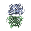
| ||||||||
| 2 | 
| ||||||||
| Unit cell |
|
-Components
| #1: Protein | Mass: 27060.859 Da / Num. of mol.: 4 Source method: isolated from a genetically manipulated source Source: (gene. exp.) Renilla reniformis (sea pansy) / Gene: GFP / Plasmid: pBAD / Production host:  #2: Water | ChemComp-HOH / |  Mass: 18.015 Da / Num. of mol.: 537 / Source method: isolated from a natural source / Formula: H2O Mass: 18.015 Da / Num. of mol.: 537 / Source method: isolated from a natural source / Formula: H2OHas protein modification | N | |
|---|
-Experimental details
-Experiment
| Experiment | Method: X-RAY DIFFRACTION / Number of used crystals: 1 |
|---|
- Sample preparation
| Crystal | Density Matthews: 2.3 Å3/Da / Density % sol: 46.63 % |
|---|---|
| Crystal grow | Temperature: 293 K / Method: vapor diffusion, hanging drop / pH: 7 Details: 2.0M ammonium sulfate, 5% v/v isopropanol, pH 7.0, VAPOR DIFFUSION, HANGING DROP, temperature 293K |
-Data collection
| Diffraction | Mean temperature: 100 K |
|---|---|
| Diffraction source | Source: SYNCHROTRON / Site: ALS  / Beamline: 8.2.2 / Beamline: 8.2.2 |
| Detector | Date: Sep 17, 2005 |
| Radiation | Protocol: SINGLE WAVELENGTH / Monochromatic (M) / Laue (L): M / Scattering type: x-ray |
| Radiation wavelength | Relative weight: 1 |
| Reflection | Resolution: 1.5→50 Å / Num. all: 123752 / Num. obs: 123752 / Observed criterion σ(F): 0 / Observed criterion σ(I): 0 |
- Processing
| Software |
| ||||||||||||||||||||
|---|---|---|---|---|---|---|---|---|---|---|---|---|---|---|---|---|---|---|---|---|---|
| Refinement | Method to determine structure: MOLECULAR REPLACEMENT Starting model: hybrid of pdb entries 1MOV, 1MOU, 1XSS and 1GGX Resolution: 1.5→50 Å / Stereochemistry target values: Engh & Huber
| ||||||||||||||||||||
| Refinement step | Cycle: LAST / Resolution: 1.5→50 Å
|
