 Movie
Movie Controller

[English] 日本語
 Yorodumi
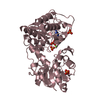
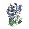
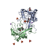
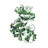
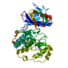
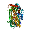
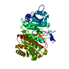
- PDB-6z2o: Crystal structure of wild type OgpA from Akkermansia muciniphila ... -
+ Open data

- Basic information
| Entry | Database: PDB / ID: 6z2o | ||||||
|---|---|---|---|---|---|---|---|
| Title | Crystal structure of wild type OgpA from Akkermansia muciniphila in P 21 21 21 | ||||||
 Components Components | O-glycan protease | ||||||
 Keywords Keywords | HYDROLASE / O-glycan endopeptidase / mucins / OgpA. metalloprotease | ||||||
| Function / homology | metal ion binding / Peptidase M43 pregnancy-associated plasma-A domain-containing protein Function and homology information Function and homology information | ||||||
| Biological species |  Akkermansia muciniphila ATCC BAA-835 (bacteria) Akkermansia muciniphila ATCC BAA-835 (bacteria) | ||||||
| Method |  X-RAY DIFFRACTION / SYNCHROTRON / MOLECULAR REPLACEMENT / Resolution: 1.649 Å X-RAY DIFFRACTION / SYNCHROTRON / MOLECULAR REPLACEMENT / Resolution: 1.649 Å | ||||||
 Authors Authors | Trastoy, B. / Naegali, A. / Anso, I. / Sjogren, J. / Guerin, M.E. | ||||||
 Citation Citation | Journal: Nat Commun / Year: 2020 Title: Structural basis of mammalian mucin processing by the human gut O-glycopeptidase OgpA from Akkermansia muciniphila. Authors: Trastoy, B. / Naegeli, A. / Anso, I. / Sjogren, J. / Guerin, M.E. | ||||||
| History |
|
- Structure visualization
| Structure viewer | Molecule: MolmilJmol/JSmol |
|---|
- Downloads & links
-Download
| PDBx/mmCIF format | 6z2o.cif.gz | 93.4 KB | Display | PDBx/mmCIF format |
|---|---|---|---|---|
| PDB format | pdb6z2o.ent.gz | 66.9 KB | Display | PDB format |
| PDBx/mmJSON format | 6z2o.json.gz | Tree view | PDBx/mmJSON format | |
| Others |  Other downloads Other downloads |
-Validation report
| Arichive directory | https://data.pdbj.org/pub/pdb/validation_reports/z2/6z2oftp://data.pdbj.org/pub/pdb/validation_reports/z2/6z2o | HTTPS FTP |
|---|
-Related structure data
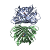
| Related structure data |  6z2dSC 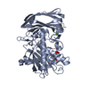 6z2pC 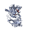 6z2qC S: Starting model for refinement C: citing same article ( |
|---|---|
| Similar structure data |
-Links
 PDBj
PDBj- Assembly
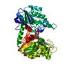
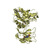
| Deposited unit | 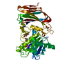
| ||||||||
|---|---|---|---|---|---|---|---|---|---|
| 1 |
| ||||||||
| Unit cell |
|
-Components
| #1: Protein | Mass: 41856.137 Da / Num. of mol.: 1 Source method: isolated from a genetically manipulated source Source: (gene. exp.) Akkermansia muciniphila ATCC BAA-835 (bacteria)Gene: Amuc_1119 / Production host: | ||||||
|---|---|---|---|---|---|---|---|
| #2: Chemical | ChemComp-EDO /   Mass: 62.068 Da / Num. of mol.: 7 / Source method: obtained synthetically / Formula: C2H6O2 Mass: 62.068 Da / Num. of mol.: 7 / Source method: obtained synthetically / Formula: C2H6O2#3: Chemical | ChemComp-ZN / |   Mass: 65.409 Da / Num. of mol.: 1 / Source method: obtained synthetically / Formula: Zn Mass: 65.409 Da / Num. of mol.: 1 / Source method: obtained synthetically / Formula: Zn#4: Water | ChemComp-HOH / |  Mass: 18.015 Da / Num. of mol.: 257 / Source method: isolated from a natural source / Formula: H2O Mass: 18.015 Da / Num. of mol.: 257 / Source method: isolated from a natural source / Formula: H2OHas ligand of interest | N | |
-Experimental details
-Experiment
| Experiment | Method: X-RAY DIFFRACTION / Number of used crystals: 1 |
|---|
- Sample preparation
| Crystal | Density Matthews: 2.6 Å3/Da / Density % sol: 52.7 % |
|---|---|
| Crystal grow | Temperature: 291 K / Method: vapor diffusion, sitting drop Details: 100mM sodium imidazole/MES monohydrate pH 6.5, 100mM carboxylic acids mixture (sodium formate, ammonium acetate, sodium citrate tribasic dihydrate, sodium potassium tartrate tetrahydrate and ...Details: 100mM sodium imidazole/MES monohydrate pH 6.5, 100mM carboxylic acids mixture (sodium formate, ammonium acetate, sodium citrate tribasic dihydrate, sodium potassium tartrate tetrahydrate and sodium oxamate) and 50% (w/v) of precipitant mix based on 25% (w/v) MPD, 25% (w/v) PEG 1000 and 25% (w/v) PEG 3350 (Morpheus protein crystallization screen). |
-Data collection
| Diffraction | Mean temperature: 100 K / Serial crystal experiment: N |
|---|---|
| Diffraction source | Source: SYNCHROTRON / Site: Diamond  / Beamline: I24 / Wavelength: 1.1702 Å / Beamline: I24 / Wavelength: 1.1702 Å |
| Detector | Type: DECTRIS PILATUS 6M / Detector: PIXEL / Date: May 16, 2019 |
| Radiation | Protocol: SINGLE WAVELENGTH / Monochromatic (M) / Laue (L): M / Scattering type: x-ray |
| Radiation wavelength | Wavelength: 1.1702 Å / Relative weight: 1 |
| Reflection | Resolution: 1.649→29.632 Å / Num. obs: 52999 / % possible obs: 99.66 % / Redundancy: 12.6 % / Biso Wilson estimate: 23.23 Å2 / CC1/2: 0.998 / CC star: 0.999 / Rmerge(I) obs: 0.08763 / Rrim(I) all: 0.09136 / Net I/σ(I): 16.95 |
| Reflection shell | Resolution: 1.649→1.708 Å / Redundancy: 10.5 % / Rmerge(I) obs: 0.5749 / Mean I/σ(I) obs: 3.29 / Num. unique obs: 5089 / CC1/2: 0.1835 / CC star: 0.905 / Rrim(I) all: 0.6045 / % possible all: 97.03 |
- Processing
| Software |
| ||||||||||||||||||||||||||||||||||||||||||||||||||||||||||||||||||||||||||||||||||||||||||||||||||||||||||||||||||||||||
|---|---|---|---|---|---|---|---|---|---|---|---|---|---|---|---|---|---|---|---|---|---|---|---|---|---|---|---|---|---|---|---|---|---|---|---|---|---|---|---|---|---|---|---|---|---|---|---|---|---|---|---|---|---|---|---|---|---|---|---|---|---|---|---|---|---|---|---|---|---|---|---|---|---|---|---|---|---|---|---|---|---|---|---|---|---|---|---|---|---|---|---|---|---|---|---|---|---|---|---|---|---|---|---|---|---|---|---|---|---|---|---|---|---|---|---|---|---|---|---|---|---|
| Refinement | Method to determine structure: MOLECULAR REPLACEMENT Starting model: 6Z2D Resolution: 1.649→29.632 Å / SU ML: 0.17 / Cross valid method: THROUGHOUT / σ(F): 1.35 / Phase error: 22.19 / Stereochemistry target values: ML
| ||||||||||||||||||||||||||||||||||||||||||||||||||||||||||||||||||||||||||||||||||||||||||||||||||||||||||||||||||||||||
| Solvent computation | Shrinkage radii: 0.9 Å / VDW probe radii: 1.11 Å / Solvent model: FLAT BULK SOLVENT MODEL | ||||||||||||||||||||||||||||||||||||||||||||||||||||||||||||||||||||||||||||||||||||||||||||||||||||||||||||||||||||||||
| Displacement parameters | Biso max: 73.31 Å2 / Biso mean: 27.9432 Å2 / Biso min: 15.42 Å2 | ||||||||||||||||||||||||||||||||||||||||||||||||||||||||||||||||||||||||||||||||||||||||||||||||||||||||||||||||||||||||
| Refinement step | Cycle: final / Resolution: 1.649→29.632 Å
| ||||||||||||||||||||||||||||||||||||||||||||||||||||||||||||||||||||||||||||||||||||||||||||||||||||||||||||||||||||||||
| LS refinement shell | Refine-ID: X-RAY DIFFRACTION / Rfactor Rfree error: 0
|
