 Movie
Movie Controller

[English] 日本語
 Yorodumi
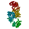
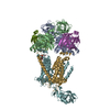
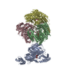
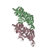
- PDB-2r82: Pyruvate phosphate dikinase (PPDK) triple mutant R219E/E271R/S262... -
+ Open data

- Basic information
| Entry | Database: PDB / ID: 2r82 | ||||||
|---|---|---|---|---|---|---|---|
| Title | Pyruvate phosphate dikinase (PPDK) triple mutant R219E/E271R/S262D adapts a second conformational state | ||||||
 Components Components | Pyruvate, phosphate dikinase | ||||||
 Keywords Keywords | TRANSFERASE / phosphotransferase / conformational transition / swiveling domain / remote active sites / ATP-binding / Kinase / Magnesium / Metal-binding / Nucleotide-binding / Phosphorylation | ||||||
| Function / homology |  Function and homology information Function and homology informationpyruvate, phosphate dikinase / pyruvate, phosphate dikinase activity / kinase activity / ATP binding / metal ion binding Similarity search - Function | ||||||
| Biological species |  Clostridium symbiosum (bacteria) Clostridium symbiosum (bacteria) | ||||||
| Method |  X-RAY DIFFRACTION / MOLECULAR REPLACEMENT / Resolution: 3.6 Å X-RAY DIFFRACTION / MOLECULAR REPLACEMENT / Resolution: 3.6 Å | ||||||
 Authors Authors | Lim, K. / Read, R.J. / Chen, C.C. / Herzberg, O. | ||||||
 Citation Citation | Journal: Biochemistry / Year: 2007 Title: Swiveling domain mechanism in pyruvate phosphate dikinase. Authors: Lim, K. / Read, R.J. / Chen, C.C. / Tempczyk, A. / Wei, M. / Ye, D. / Wu, C. / Dunaway-Mariano, D. / Herzberg, O. #1: Journal: Biochemistry / Year: 2002Title: Pyruvate site of pyruvate phosphate dikinase: crystal structure of the enzyme-phosphonopyruvate complex, and mutant analysis Authors: Herzberg, O. / Chen, C. / Liu, S. / Tempczyk, A. / Howard, A. / Wei, M. / Ye, D. / Dunaway-Mariano, D. #2: Journal: Proc.Natl.Acad.Sci.USA / Year: 1996Title: Swiveling-domain mechanism for enzymatic phosphotransfer between remote reaction sites Authors: Herzberg, O. / Chen, C. / Kapadia, G. / Mcguire, M. / Carroll, L.J. / Noh, S.J. / Dunaway-Mariano, D. #3: Journal: Biochemistry / Year: 1998Title: Location of the phosphate binding site within Clostridium Symbiosum pyruvate phosphate dikinase Authors: Mcguire, M. / Huang, K. / Kapadia, G. / Herzberg, O. / Dunaway-Mariano, D. #4: Journal: J.Biol.Chem. / Year: 2000Title: Identification of the domain-domain docking sites within Clostridium Symbiosum pyruvate phosphate dikinase by amino acid replacement Authors: Wei, M. / Li, Z. / Ye, D. / Herzberg, O. / Dunaway-Mariano, D. | ||||||
| History |
|
- Structure visualization
| Structure viewer | Molecule: MolmilJmol/JSmol |
|---|
- Downloads & links
-Download
| PDBx/mmCIF format | 2r82.cif.gz | 162.3 KB | Display | PDBx/mmCIF format |
|---|---|---|---|---|
| PDB format | pdb2r82.ent.gz | 129 KB | Display | PDB format |
| PDBx/mmJSON format | 2r82.json.gz | Tree view | PDBx/mmJSON format | |
| Others |  Other downloads Other downloads |
-Validation report
| Arichive directory | https://data.pdbj.org/pub/pdb/validation_reports/r8/2r82ftp://data.pdbj.org/pub/pdb/validation_reports/r8/2r82 | HTTPS FTP |
|---|
-Related structure data
| Related structure data |  9pzlC  1kblS S: Starting model for refinement C: citing same article ( |
|---|---|
| Similar structure data |


-Links
 PDBj
PDBj


- Assembly
| Deposited unit | 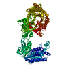
| ||||||||
|---|---|---|---|---|---|---|---|---|---|
| 1 | 
| ||||||||
| Unit cell |
|
-Components
| #1: Protein | Mass: 96799.359 Da / Num. of mol.: 1 / Mutation: R219E, S262D, E271R Source method: isolated from a genetically manipulated source Source: (gene. exp.) Clostridium symbiosum (bacteria) / Gene: ppdK / Plasmid: pACYC184 D12 / Production host: |
|---|---|
| #2: Chemical |   Mass: 96.063 Da / Num. of mol.: 2 / Source method: obtained synthetically / Formula: SO4 Mass: 96.063 Da / Num. of mol.: 2 / Source method: obtained synthetically / Formula: SO4 |
-Experimental details
-Experiment
| Experiment | Method: X-RAY DIFFRACTION / Number of used crystals: 1 |
|---|
- Sample preparation
| Crystal | Density Matthews: 2.69 Å3/Da / Density % sol: 54.19 % |
|---|---|
| Crystal grow | Temperature: 303 K / Method: vapor diffusion, hanging drop / pH: 7 Details: 50% saturated ammonium sulfate, 0.1 M Na Hepes, 28 mg/ml protein (in 20 mM imidazole (pH 6.5), 0.1 mM EDTA, 100 mM KCl, and 1mM DTT), pH 7.0, VAPOR DIFFUSION, HANGING DROP, temperature 303K |
-Data collection
| Diffraction | Mean temperature: 100 K |
|---|---|
| Diffraction source | Source: ROTATING ANODE / Type: SIEMENS / Wavelength: 1.5418 Å |
| Detector | Type: MAR scanner 345 mm plate / Detector: IMAGE PLATE |
| Radiation | Monochromator: Harvard mirrors / Protocol: SINGLE WAVELENGTH / Monochromatic (M) / Laue (L): M / Scattering type: x-ray |
| Radiation wavelength | Wavelength: 1.5418 Å / Relative weight: 1 |
| Reflection | Resolution: 3.6→50 Å / Num. all: 11406 / Num. obs: 11406 / % possible obs: 91 % / Observed criterion σ(F): 0 / Observed criterion σ(I): 0 / Redundancy: 3.6 % / Rmerge(I) obs: 0.155 / Net I/σ(I): 6.1 |
| Reflection shell | Resolution: 3.6→3.76 Å / Redundancy: 2 % / Rmerge(I) obs: 0.331 / Mean I/σ(I) obs: 2.5 / % possible all: 76.5 |
- Processing
| Software |
| |||||||||||||||||||||||||
|---|---|---|---|---|---|---|---|---|---|---|---|---|---|---|---|---|---|---|---|---|---|---|---|---|---|---|
| Refinement | Method to determine structure: MOLECULAR REPLACEMENT Starting model: PDB entry 1KBL Resolution: 3.6→50 Å / Isotropic thermal model: isotropic / Cross valid method: THROUGHOUT / σ(F): 0 / σ(I): 0 / Stereochemistry target values: Engh & Huber
| |||||||||||||||||||||||||
| Displacement parameters | Biso mean: 68 Å2 | |||||||||||||||||||||||||
| Refinement step | Cycle: LAST / Resolution: 3.6→50 Å
| |||||||||||||||||||||||||
| Refine LS restraints |
| |||||||||||||||||||||||||
| LS refinement shell | Resolution: 3.6→3.76 Å / Rfactor Rfree: 0.38 / Rfactor Rwork: 0.35 |
