 Movie
Movie Controller


+ Open data

- Basic information
| Entry | Database: PDB / ID: 1gl9 | ||||||
|---|---|---|---|---|---|---|---|
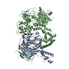
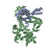
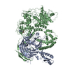
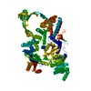
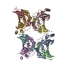
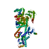
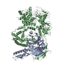
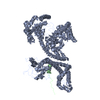
| Title | Archaeoglobus fulgidus reverse gyrase complexed with ADPNP | ||||||
 Components Components | REVERSE GYRASE | ||||||
 Keywords Keywords | TOPOISOMERASE / DNA SUPERCOILING / ARCHAEA / HELICASE | ||||||
| Function / homology |  Function and homology information Function and homology informationreverse gyrase activity / Isomerases; Isomerases altering macromolecular conformation; Enzymes altering nucleic acid conformation / DNA topological change / ATP-dependent activity, acting on DNA / DNA replication / ATP hydrolysis activity / DNA binding / zinc ion binding / ATP binding / cytoplasm Similarity search - Function | ||||||
| Biological species |   ARCHAEOGLOBUS FULGIDUS (archaea) ARCHAEOGLOBUS FULGIDUS (archaea) | ||||||
| Method |  X-RAY DIFFRACTION / SYNCHROTRON / MOLECULAR REPLACEMENT / Resolution: 3.2 Å X-RAY DIFFRACTION / SYNCHROTRON / MOLECULAR REPLACEMENT / Resolution: 3.2 Å | ||||||
 Authors Authors | Rodriguez, A.C. / Stock, D. | ||||||
 Citation Citation | Journal: Embo J. / Year: 2002 Title: Crystal Structure of Reverse Gyrase: Insights Into the Positive Supercoiling of DNA. Authors: Rodriguez, A.C. / Stock, D. | ||||||
| History |
| ||||||
| Remark 700 | SHEET THE SHEET STRUCTURE OF THIS MOLECULE IS BIFURCATED. IN ORDER TO REPRESENT THIS FEATURE IN ... SHEET THE SHEET STRUCTURE OF THIS MOLECULE IS BIFURCATED. IN ORDER TO REPRESENT THIS FEATURE IN THE SHEET RECORDS BELOW, TWO SHEETS ARE DEFINED. |
- Structure visualization
| Structure viewer | Molecule: MolmilJmol/JSmol |
|---|
- Downloads & links
-Download
| PDBx/mmCIF format | 1gl9.cif.gz | 419 KB | Display | PDBx/mmCIF format |
|---|---|---|---|---|
| PDB format | pdb1gl9.ent.gz | 335.1 KB | Display | PDB format |
| PDBx/mmJSON format | 1gl9.json.gz | Tree view | PDBx/mmJSON format | |
| Others |  Other downloads Other downloads |
-Validation report
| Arichive directory | https://data.pdbj.org/pub/pdb/validation_reports/gl/1gl9ftp://data.pdbj.org/pub/pdb/validation_reports/gl/1gl9 | HTTPS FTP |
|---|
-Related structure data
| Related structure data |  1gkuSC S: Starting model for refinement C: citing same article ( |
|---|---|
| Similar structure data |


-Links
 PDBj
PDBj


- Assembly
| Deposited unit | 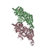
| ||||||||
|---|---|---|---|---|---|---|---|---|---|
| 1 | 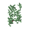
| ||||||||
| 2 | 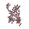
| ||||||||
| Unit cell |
| ||||||||
| Noncrystallographic symmetry (NCS) | NCS oper: (Code: given Matrix: (0.98909, -0.06165, -0.13379), Vector: |
-Components
| #1: Protein | Mass: 121535.023 Da / Num. of mol.: 2 / Mutation: YES Source method: isolated from a genetically manipulated source Details: ADENYLYLIMIDODIPHOSPHATE (ADPNP) / Source: (gene. exp.) ARCHAEOGLOBUS FULGIDUS (archaea) / Strain: VC-16 / Description: GERMAN COLLECTION OF MICROORGANISMS (DSMZ) / Plasmid: PRET3A / Production host:  #2: Chemical |   Mass: 506.196 Da / Num. of mol.: 2 / Source method: obtained synthetically / Formula: C10H17N6O12P3 / Comment: AMP-PNP, energy-carrying molecule analogue*YM Mass: 506.196 Da / Num. of mol.: 2 / Source method: obtained synthetically / Formula: C10H17N6O12P3 / Comment: AMP-PNP, energy-carrying molecule analogue*YM#3: Chemical |   Mass: 24.305 Da / Num. of mol.: 2 / Source method: obtained synthetically / Formula: Mg Mass: 24.305 Da / Num. of mol.: 2 / Source method: obtained synthetically / Formula: MgCompound details | CHAIN B ENGINEERED MUTATION PRO719LEU, LEU1046MET. RESIDUES N-TERMINAL TO B31 IN MOLECULE B, AND TO ...CHAIN B ENGINEERED | Has protein modification | Y | |
|---|
-Experimental details
-Experiment
| Experiment | Method: X-RAY DIFFRACTION / Number of used crystals: 1 |
|---|
- Sample preparation
| Crystal | Density Matthews: 2.44 Å3/Da / Density % sol: 58.7 % | |||||||||||||||||||||||||||||||||||||||||||||||||||||||||||||||
|---|---|---|---|---|---|---|---|---|---|---|---|---|---|---|---|---|---|---|---|---|---|---|---|---|---|---|---|---|---|---|---|---|---|---|---|---|---|---|---|---|---|---|---|---|---|---|---|---|---|---|---|---|---|---|---|---|---|---|---|---|---|---|---|---|
| Crystal grow | pH: 6 Details: 15% PEG 1000, 15% ETHYLENE GLYCOL 100 MM CACODYLATE (PH 6),2MM ADPNP | |||||||||||||||||||||||||||||||||||||||||||||||||||||||||||||||
| Crystal grow | *PLUS Temperature: 18 ℃ / pH: 8 / Method: vapor diffusion, sitting drop | |||||||||||||||||||||||||||||||||||||||||||||||||||||||||||||||
| Components of the solutions | *PLUS
|
-Data collection
| Diffraction | Mean temperature: 100 K |
|---|---|
| Diffraction source | Source: SYNCHROTRON / Site: ESRF  / Beamline: ID14-4 / Wavelength: 0.9686 / Beamline: ID14-4 / Wavelength: 0.9686 |
| Detector | Type: ADSC / Date: Feb 15, 2001 |
| Radiation | Protocol: SINGLE WAVELENGTH / Monochromatic (M) / Laue (L): M / Scattering type: x-ray |
| Radiation wavelength | Wavelength: 0.9686 Å / Relative weight: 1 |
| Reflection | Resolution: 3.2→34 Å / Num. obs: 38400 / % possible obs: 96.7 % / Redundancy: 3.3 % / Biso Wilson estimate: 81 Å2 / Rsym value: 0.102 / Net I/σ(I): 5.2 |
| Reflection shell | Resolution: 3.2→3.35 Å / Redundancy: 2.9 % / Mean I/σ(I) obs: 2.3 / Rsym value: 0.321 / % possible all: 96.7 |
| Reflection | *PLUS Lowest resolution: 34 Å / Num. obs: 38401 / Rmerge(I) obs: 0.102 |
| Reflection shell | *PLUS % possible obs: 96.7 % / Rmerge(I) obs: 0.321 |
- Processing
| Software |
| ||||||||||||||||||||||||||||||||||||||||||||||||||||||||||||
|---|---|---|---|---|---|---|---|---|---|---|---|---|---|---|---|---|---|---|---|---|---|---|---|---|---|---|---|---|---|---|---|---|---|---|---|---|---|---|---|---|---|---|---|---|---|---|---|---|---|---|---|---|---|---|---|---|---|---|---|---|---|
| Refinement | Method to determine structure: MOLECULAR REPLACEMENT Starting model: PDB ENTRY 1GKU Resolution: 3.2→34 Å / Data cutoff high absF: 10000 / Cross valid method: THROUGHOUT / σ(F): 0 / Stereochemistry target values: MLF Details: RESIDUES N-TERMINAL TO B31 IN MOLECULE B, AND TO RESIDUE C32 IN MOLECULE C HAVE BEEN MODELLED AS POLYALANINE. THEIR IDENTITY AND NUMBERING IN THE PROTEIN SEQUENCE IS ARBITRARY.
| ||||||||||||||||||||||||||||||||||||||||||||||||||||||||||||
| Solvent computation | Bsol: 59.5563 Å2 / ksol: 0.285991 e/Å3 | ||||||||||||||||||||||||||||||||||||||||||||||||||||||||||||
| Displacement parameters | Biso mean: 75.6 Å2
| ||||||||||||||||||||||||||||||||||||||||||||||||||||||||||||
| Refine analyze |
| ||||||||||||||||||||||||||||||||||||||||||||||||||||||||||||
| Refinement step | Cycle: LAST / Resolution: 3.2→34 Å
| ||||||||||||||||||||||||||||||||||||||||||||||||||||||||||||
| Refine LS restraints |
| ||||||||||||||||||||||||||||||||||||||||||||||||||||||||||||
| Refine LS restraints NCS | Weight Biso : 10 / Weight position: 15 | ||||||||||||||||||||||||||||||||||||||||||||||||||||||||||||
| LS refinement shell | Resolution: 3.2→3.31 Å / Total num. of bins used: 10
| ||||||||||||||||||||||||||||||||||||||||||||||||||||||||||||
| Xplor file |
| ||||||||||||||||||||||||||||||||||||||||||||||||||||||||||||
| Software | *PLUS Name: CNS / Version: 1.1 / Classification: refinement | ||||||||||||||||||||||||||||||||||||||||||||||||||||||||||||
| Refinement | *PLUS Lowest resolution: 34 Å | ||||||||||||||||||||||||||||||||||||||||||||||||||||||||||||
| Solvent computation | *PLUS | ||||||||||||||||||||||||||||||||||||||||||||||||||||||||||||
| Displacement parameters | *PLUS |
