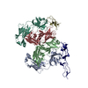
SHEET THE SHEET STRUCTURE OF THIS MOLECULE IS BIFURCATED. IN ORDER TO REPRESENT THIS FEATURE IN ... SHEET THE SHEET STRUCTURE OF THIS MOLECULE IS BIFURCATED. IN ORDER TO REPRESENT THIS FEATURE IN THE SHEET RECORDS BELOW, TWO SHEETS ARE DEFINED.
Mass: 18.015 Da / Num. of mol.: 157 / Source method: isolated from a natural source / Formula: H2O
Compound details
CHAIN B ENGINEERED MUTATION PRO719LEU, LEU1046MET. NINETEEN RESIDUES N-TERMINAL TO SERINE 33 IN THE ...CHAIN B ENGINEERED MUTATION PRO719LEU, LEU1046MET. NINETEEN RESIDUES N-TERMINAL TO SERINE 33 IN THE COORDINATES HAVE BEEN MODELLED AS THREE NON-CONTIGUOUS CHAINS OF POLYALANINE. AMINO ACID SEQUENCE AND NUMBERING FOR THESE RESIDUES HAS BEEN ASSIGNED AS 1-21. THEIR ACTUAL POSITION IN THE PROTEIN SEQUENCE IS UNCLEAR DUE TO POOR ELECTRON DENSITY.
Has protein modification
Y
-
Experimental details
-
Experiment
Experiment
Method: X-RAY DIFFRACTION / Number of used crystals: 1
-
Sample preparation
Crystal
Density Matthews: 2.26 Å3/Da / Density % sol: 52 %
Protocol: SINGLE WAVELENGTH / Monochromatic (M) / Laue (L): M / Scattering type: x-ray
Radiation wavelength
Wavelength: 0.9686 Å / Relative weight: 1
Reflection
Resolution: 2.7→41 Å / Num. obs: 28735 / % possible obs: 99.3 % / Redundancy: 3.2 % / Biso Wilson estimate: 67.6 Å2 / Rsym value: 0.062 / Net I/σ(I): 8.6
Reflection shell
Resolution: 2.7→2.78 Å / Redundancy: 3 % / Mean I/σ(I) obs: 2.6 / Rsym value: 0.288 / % possible all: 99.3
Reflection
*PLUS
Lowest resolution: 41 Å / Rmerge(I) obs: 0.062
Reflection shell
*PLUS
% possible obs: 99.3 % / Rmerge(I) obs: 0.288
-
Processing
Software
Name
Version
Classification
CNS
1
refinement
MOSFLM
datareduction
SCALA
datascaling
SOLVE/RESOLVE
phasing
Refinement
Method to determine structure: MAD / Resolution: 2.7→41 Å / Data cutoff high absF: 10000 / Cross valid method: THROUGHOUT / σ(F): 0 / Stereochemistry target values: MLF Details: NINETEEN RESIDUES N-TERMINAL TO SERINE 33 IN THE COORDINATES HAVE BEEN MODELLED AS THREE NON- CONTIGUOUS CHAINS OF POLYALANINE. AMINO ACID SEQUENCE AND NUMBERING FOR THESE RESIDUES HAS BEEN ...Details: NINETEEN RESIDUES N-TERMINAL TO SERINE 33 IN THE COORDINATES HAVE BEEN MODELLED AS THREE NON- CONTIGUOUS CHAINS OF POLYALANINE. AMINO ACID SEQUENCE AND NUMBERING FOR THESE RESIDUES HAS BEEN ASSIGNED AS 1-21. THEIR ACTUAL POSITION IN THE PROTEIN SEQUENCE IS UNCLEAR DUE TO POOR ELECTRON DENSITY.
Rfactor
Num. reflection
% reflection
Selection details
Rfree
0.295
1474
4.8 %
RANDOM
Rwork
0.226
-
-
-
obs
0.226
30333
99.3 %
-
Solvent computation
Bsol: 66.5478 Å2 / ksol: 0.35335 e/Å3
Displacement parameters
Biso mean: 55.4 Å2
Baniso -1
Baniso -2
Baniso -3
1-
7.13 Å2
0 Å2
0.438 Å2
2-
-
-7.426 Å2
0 Å2
3-
-
-
0.296 Å2
Refine analyze
Free
Obs
Luzzati coordinate error
0.49 Å
0.34 Å
Luzzati d res low
-
5 Å
Luzzati sigma a
0.45 Å
0.23 Å
Refinement step
Cycle: LAST / Resolution: 2.7→41 Å
Protein
Nucleic acid
Ligand
Solvent
Total
Num. atoms
8151
0
0
157
8308
Refine LS restraints
Refine-ID
Type
Dev ideal
Dev ideal target
X-RAY DIFFRACTION
c_bond_d
0.007
X-RAY DIFFRACTION
c_bond_d_na
X-RAY DIFFRACTION
c_bond_d_prot
X-RAY DIFFRACTION
c_angle_d
X-RAY DIFFRACTION
c_angle_d_na
X-RAY DIFFRACTION
c_angle_d_prot
X-RAY DIFFRACTION
c_angle_deg
1.3
X-RAY DIFFRACTION
c_angle_deg_na
X-RAY DIFFRACTION
c_angle_deg_prot
X-RAY DIFFRACTION
c_dihedral_angle_d
X-RAY DIFFRACTION
c_dihedral_angle_d_na
X-RAY DIFFRACTION
c_dihedral_angle_d_prot
X-RAY DIFFRACTION
c_improper_angle_d
X-RAY DIFFRACTION
c_improper_angle_d_na
X-RAY DIFFRACTION
c_improper_angle_d_prot
X-RAY DIFFRACTION
c_mcbond_it
3.42
3
X-RAY DIFFRACTION
c_mcangle_it
5.77
4
X-RAY DIFFRACTION
c_scbond_it
5.28
4
X-RAY DIFFRACTION
c_scangle_it
8.11
6
LS refinement shell
Resolution: 2.7→2.8 Å / Total num. of bins used: 10
In the structure databanks used in Yorodumi, some data are registered as the other names, "COVID-19 virus" and "2019-nCoV". Here are the details of the virus and the list of structure data.
Jan 31, 2019. EMDB accession codes are about to change! (news from PDBe EMDB page)
EMDB accession codes are about to change! (news from PDBe EMDB page)
The allocation of 4 digits for EMDB accession codes will soon come to an end. Whilst these codes will remain in use, new EMDB accession codes will include an additional digit and will expand incrementally as the available range of codes is exhausted. The current 4-digit format prefixed with “EMD-” (i.e. EMD-XXXX) will advance to a 5-digit format (i.e. EMD-XXXXX), and so on. It is currently estimated that the 4-digit codes will be depleted around Spring 2019, at which point the 5-digit format will come into force.
The EM Navigator/Yorodumi systems omit the EMD- prefix.
Related info.:Q: What is EMD? / ID/Accession-code notation in Yorodumi/EM Navigator
Yorodumi is a browser for structure data from EMDB, PDB, SASBDB, etc.
This page is also the successor to EM Navigator detail page, and also detail information page/front-end page for Omokage search.
The word "yorodu" (or yorozu) is an old Japanese word meaning "ten thousand". "mi" (miru) is to see.
Related info.:EMDB / PDB / SASBDB / Comparison of 3 databanks / Yorodumi Search / Aug 31, 2016. New EM Navigator & Yorodumi / Yorodumi Papers / Jmol/JSmol / Function and homology information / Changes in new EM Navigator and Yorodumi
 Movie
Movie Controller


 Open data
 Basic information
 Components
 Keywords
 Function and homology information
 ARCHAEOGLOBUS FULGIDUS (archaea)
 X-RAY DIFFRACTION /
  Authors
 Citation
 Structure visualization
 Downloads & links
 Other downloads


 PDBj
PDBj

 Assembly

 Mass: 18.015 Da / Num. of mol.: 157 / Source method: isolated from a natural source / Formula: H2O
 Sample preparation
 / Beamline: ID14-4 / Wavelength: 0.9686
  Processing