- PDB-2r4v: Structure of human CLIC2, crystal form A -
+
Open data
ID or keywords:
Loading...
-
Basic information
Entry
Database: PDB / ID: 2r4v
Title
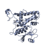
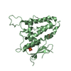
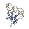
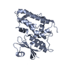
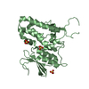
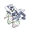
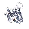
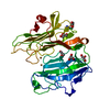
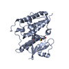
Structure of human CLIC2, crystal form A
Components
Chloride intracellular channel protein 2
Keywords
TRANSPORT PROTEIN / chloride intracellular channels / CLIC2 / pore-forming protein / ryanodine receptor / Chloride channel / Ion transport / Ionic channel / Transport / Voltage-gated channel
Function / homology
Function and homology information
glutathione peroxidase activity / Oxidoreductases; Acting on a sulfur group of donors / chloride transport / chloride channel activity / Oxidoreductases; Acting on a peroxide as acceptor; Peroxidases / chloride channel complex / regulation of release of sequestered calcium ion into cytosol by sarcoplasmic reticulum / Ion homeostasis / regulation of cardiac muscle contraction by regulation of the release of sequestered calcium ion / Stimuli-sensing channels ...glutathione peroxidase activity / Oxidoreductases; Acting on a sulfur group of donors / chloride transport / chloride channel activity / Oxidoreductases; Acting on a peroxide as acceptor; Peroxidases / chloride channel complex / regulation of release of sequestered calcium ion into cytosol by sarcoplasmic reticulum / Ion homeostasis / regulation of cardiac muscle contraction by regulation of the release of sequestered calcium ion / Stimuli-sensing channels / signal transduction / membrane / nucleus / plasma membrane / cytoplasm / cytosol Similarity search - Function
In the structure databanks used in Yorodumi, some data are registered as the other names, "COVID-19 virus" and "2019-nCoV". Here are the details of the virus and the list of structure data.
Jan 31, 2019. EMDB accession codes are about to change! (news from PDBe EMDB page)
EMDB accession codes are about to change! (news from PDBe EMDB page)
The allocation of 4 digits for EMDB accession codes will soon come to an end. Whilst these codes will remain in use, new EMDB accession codes will include an additional digit and will expand incrementally as the available range of codes is exhausted. The current 4-digit format prefixed with “EMD-” (i.e. EMD-XXXX) will advance to a 5-digit format (i.e. EMD-XXXXX), and so on. It is currently estimated that the 4-digit codes will be depleted around Spring 2019, at which point the 5-digit format will come into force.
The EM Navigator/Yorodumi systems omit the EMD- prefix.
Related info.:Q: What is EMD? / ID/Accession-code notation in Yorodumi/EM Navigator
Yorodumi is a browser for structure data from EMDB, PDB, SASBDB, etc.
This page is also the successor to EM Navigator detail page, and also detail information page/front-end page for Omokage search.
The word "yorodu" (or yorozu) is an old Japanese word meaning "ten thousand". "mi" (miru) is to see.
Related info.:EMDB / PDB / SASBDB / Comparison of 3 databanks / Yorodumi Search / Aug 31, 2016. New EM Navigator & Yorodumi / Yorodumi Papers / Jmol/JSmol / Function and homology information / Changes in new EM Navigator and Yorodumi
 Movie
Movie Controller


 Open data
 Basic information
 Components
 Keywords
 Function and homology information
 Homo sapiens (human)
 X-RAY DIFFRACTION /
  Authors
 Citation
 Structure visualization
 Downloads & links
 Other downloads


 PDBj
PDBj


 Assembly
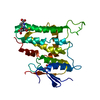


 Mass: 307.323 Da / Num. of mol.: 2 / Source method: obtained synthetically / Formula: C10H17N3O6S
 Mass: 18.015 Da / Num. of mol.: 125 / Source method: isolated from a natural source / Formula: H2O
 Sample preparation
 / Beamline: 14-ID-B / Wavelength: 0.9 Å
 Processing