 Movie
Movie Controller

[English] 日本語
 Yorodumi
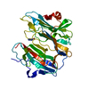
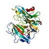
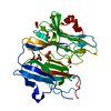
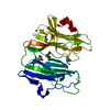
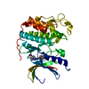
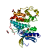
- PDB-3phm: REDUCED (CU+) PEPTIDYLGLYCINE ALPHA-HYDROXYLATING MONOOXYGENASE (PHM) -
+ Open data

- Basic information
| Entry | Database: PDB / ID: 3phm | ||||||
|---|---|---|---|---|---|---|---|
| Title | REDUCED (CU+) PEPTIDYLGLYCINE ALPHA-HYDROXYLATING MONOOXYGENASE (PHM) | ||||||
 Components Components | PROTEIN (PEPTIDYLGLYCINE ALPHA-HYDROXYLATING MONOOXYGENASE) | ||||||
 Keywords Keywords | OXIDOREDUCTASE / MONOOXYGENASE / BIOACTIVE PEPTIDE ACTIVATION / ASCORBATE | ||||||
| Function / homology |  Function and homology information Function and homology informationpeptidylglycine monooxygenase / peptidylamidoglycolate lyase / peptide amidation / peptidylglycine monooxygenase activity / peptidylamidoglycolate lyase activity / fatty acid primary amide biosynthetic process / ovulation cycle process / toxin metabolic process / long-chain fatty acid metabolic process / peptide metabolic process ...peptidylglycine monooxygenase / peptidylamidoglycolate lyase / peptide amidation / peptidylglycine monooxygenase activity / peptidylamidoglycolate lyase activity / fatty acid primary amide biosynthetic process / ovulation cycle process / toxin metabolic process / long-chain fatty acid metabolic process / peptide metabolic process / mitotic chromosome condensation / response to pH / L-ascorbic acid binding / response to zinc ion / response to copper ion / transport vesicle membrane / limb development / maternal process involved in female pregnancy / condensed chromosome / lactation / secretory granule / response to glucocorticoid / regulation of actin cytoskeleton organization / trans-Golgi network / response to estradiol / heart development / cellular response to oxidative stress / perikaryon / proteasome-mediated ubiquitin-dependent protein catabolic process / response to hypoxia / response to xenobiotic stimulus / copper ion binding / neuronal cell body / calcium ion binding / chromatin binding / regulation of transcription by RNA polymerase II / protein kinase binding / chromatin / perinuclear region of cytoplasm / cell surface / : / extracellular region / zinc ion binding / identical protein binding Similarity search - Function | ||||||
| Biological species |  | ||||||
| Method |  X-RAY DIFFRACTION / SYNCHROTRON / MOLECULAR REPLACEMENT / Resolution: 2.1 Å X-RAY DIFFRACTION / SYNCHROTRON / MOLECULAR REPLACEMENT / Resolution: 2.1 Å | ||||||
 Authors Authors | Prigge, S.T. / Amzel, L.M. | ||||||
 Citation Citation | Journal: Nat.Struct.Biol. / Year: 1999 Title: Substrate-mediated electron transfer in peptidylglycine alpha-hydroxylating monooxygenase. Authors: Prigge, S.T. / Kolhekar, A.S. / Eipper, B.A. / Mains, R.E. / Amzel, L.M. | ||||||
| History |
|
- Structure visualization
| Structure viewer | Molecule: MolmilJmol/JSmol |
|---|
- Downloads & links
-Download
| PDBx/mmCIF format | 3phm.cif.gz | 79.4 KB | Display | PDBx/mmCIF format |
|---|---|---|---|---|
| PDB format | pdb3phm.ent.gz | 58.9 KB | Display | PDB format |
| PDBx/mmJSON format | 3phm.json.gz | Tree view | PDBx/mmJSON format | |
| Others |  Other downloads Other downloads |
-Validation report
| Arichive directory | https://data.pdbj.org/pub/pdb/validation_reports/ph/3phmftp://data.pdbj.org/pub/pdb/validation_reports/ph/3phm | HTTPS FTP |
|---|
-Related structure data
| Related structure data | 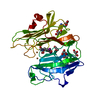 1opmC 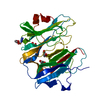 1phmS S: Starting model for refinement C: citing same article ( |
|---|---|
| Similar structure data |


-Links
 PDBj
PDBj


- Assembly
| Deposited unit | 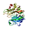
| ||||||||
|---|---|---|---|---|---|---|---|---|---|
| 1 |
| ||||||||
| Unit cell |
|
-Components
-Protein , 1 types, 1 molecules A
| #1: Protein | Mass: 34577.641 Da / Num. of mol.: 1 Source method: isolated from a genetically manipulated source Details: REDUCED (CU+), ASCORBATE SOAKED / Source: (gene. exp.)  Cricetulus griseus (Chinese hamster) / References: UniProt: P14925, peptidylglycine monooxygenase Cricetulus griseus (Chinese hamster) / References: UniProt: P14925, peptidylglycine monooxygenase |
|---|
-Non-polymers , 5 types, 177 molecules 


| #2: Chemical |  Mass: 63.546 Da / Num. of mol.: 2 / Source method: obtained synthetically / Formula: Cu Mass: 63.546 Da / Num. of mol.: 2 / Source method: obtained synthetically / Formula: Cu#3: Chemical | ChemComp-AZI / |  Mass: 42.020 Da / Num. of mol.: 1 / Source method: obtained synthetically / Formula: N3 Mass: 42.020 Da / Num. of mol.: 1 / Source method: obtained synthetically / Formula: N3#4: Chemical | ChemComp-NI / |  Mass: 58.693 Da / Num. of mol.: 1 / Source method: obtained synthetically / Formula: Ni Mass: 58.693 Da / Num. of mol.: 1 / Source method: obtained synthetically / Formula: Ni#5: Chemical | ChemComp-GOL /  Mass: 92.094 Da / Num. of mol.: 5 / Source method: obtained synthetically / Formula: C3H8O3 Mass: 92.094 Da / Num. of mol.: 5 / Source method: obtained synthetically / Formula: C3H8O3#6: Water | ChemComp-HOH / | Mass: 18.015 Da / Num. of mol.: 168 / Source method: isolated from a natural source / Formula: H2O |
|---|
-Details
| Has protein modification | Y |
|---|---|
| Nonpolymer details | CU 357 IS ACTIVE SITE COPPER CUA 1CU 358 IS ACTIVE SITE COPPER CUB NI1 359 FORMS A CRYSTAL CONTACT |
-Experimental details
-Experiment
| Experiment | Method: X-RAY DIFFRACTION / Number of used crystals: 1 |
|---|
- Sample preparation
| Crystal | Density Matthews: 2.76 Å3/Da / Density % sol: 63 % | |||||||||||||||||||||||||||||||||||
|---|---|---|---|---|---|---|---|---|---|---|---|---|---|---|---|---|---|---|---|---|---|---|---|---|---|---|---|---|---|---|---|---|---|---|---|---|
| Crystal grow | pH: 5.5 / Details: pH 5.5 | |||||||||||||||||||||||||||||||||||
| Crystal grow | *PLUS Temperature: 293 K / Method: vapor diffusion, hanging drop | |||||||||||||||||||||||||||||||||||
| Components of the solutions | *PLUS
|
-Data collection
| Diffraction | Mean temperature: 100 K |
|---|---|
| Diffraction source | Source: SYNCHROTRON / Site: NSLS  / Beamline: X4A / Wavelength: 0.9748 / Beamline: X4A / Wavelength: 0.9748 |
| Detector | Type: RIGAKU RAXIS IV / Detector: IMAGE PLATE / Date: Jan 1, 1998 / Details: SPHERICAL RH COATED MIRROR |
| Radiation | Monochromator: SAGITTALLY FOCUSED SI(111) / Protocol: SINGLE WAVELENGTH / Monochromatic (M) / Laue (L): M / Scattering type: x-ray |
| Radiation wavelength | Wavelength: 0.9748 Å / Relative weight: 1 |
| Reflection | Resolution: 2.1→20 Å / Num. obs: 23704 / % possible obs: 99.9 % / Observed criterion σ(I): -3 / Redundancy: 6 % / Rsym value: 0.065 / Net I/σ(I): 14.8 |
| Reflection shell | Resolution: 2.1→2.175 Å / Redundancy: 5 % / Mean I/σ(I) obs: 3.3 / Rsym value: 0.575 / % possible all: 99.6 |
| Reflection | *PLUS Num. measured all: 141184 / Rmerge(I) obs: 0.065 |
- Processing
| Software |
| ||||||||||||||||||||||||||||||||||||||||||||||||||||||||||||||||||||||||||||||||
|---|---|---|---|---|---|---|---|---|---|---|---|---|---|---|---|---|---|---|---|---|---|---|---|---|---|---|---|---|---|---|---|---|---|---|---|---|---|---|---|---|---|---|---|---|---|---|---|---|---|---|---|---|---|---|---|---|---|---|---|---|---|---|---|---|---|---|---|---|---|---|---|---|---|---|---|---|---|---|---|---|---|
| Refinement | Method to determine structure: MOLECULAR REPLACEMENT Starting model: 1PHM Resolution: 2.1→10 Å / Rfactor Rfree error: 0.0079 / Data cutoff high absF: 100000 / Data cutoff low absF: 0.1 / Isotropic thermal model: RESTRAINED / Cross valid method: THROUGHOUT / σ(F): 2
| ||||||||||||||||||||||||||||||||||||||||||||||||||||||||||||||||||||||||||||||||
| Displacement parameters |
| ||||||||||||||||||||||||||||||||||||||||||||||||||||||||||||||||||||||||||||||||
| Refinement step | Cycle: LAST / Resolution: 2.1→10 Å
| ||||||||||||||||||||||||||||||||||||||||||||||||||||||||||||||||||||||||||||||||
| Refine LS restraints |
| ||||||||||||||||||||||||||||||||||||||||||||||||||||||||||||||||||||||||||||||||
| LS refinement shell | Resolution: 2.1→2.16 Å / Rfactor Rfree error: 0.046 / Total num. of bins used: 12
| ||||||||||||||||||||||||||||||||||||||||||||||||||||||||||||||||||||||||||||||||
| Xplor file |
| ||||||||||||||||||||||||||||||||||||||||||||||||||||||||||||||||||||||||||||||||
| Software | *PLUS Name: X-PLOR / Version: 3.8 / Classification: refinement | ||||||||||||||||||||||||||||||||||||||||||||||||||||||||||||||||||||||||||||||||
| Refinement | *PLUS Highest resolution: 2.1 Å / Lowest resolution: 10 Å / σ(F): 2 / % reflection Rfree: 5 % / Rfactor obs: 0.2 / Rfactor Rwork: 0.2 | ||||||||||||||||||||||||||||||||||||||||||||||||||||||||||||||||||||||||||||||||
| Solvent computation | *PLUS | ||||||||||||||||||||||||||||||||||||||||||||||||||||||||||||||||||||||||||||||||
| Displacement parameters | *PLUS | ||||||||||||||||||||||||||||||||||||||||||||||||||||||||||||||||||||||||||||||||
| Refine LS restraints | *PLUS
| ||||||||||||||||||||||||||||||||||||||||||||||||||||||||||||||||||||||||||||||||
| LS refinement shell | *PLUS Highest resolution: 2.1 Å / Rfactor Rfree: 0.335 / % reflection Rfree: 2.8 % / Rfactor Rwork: 0.261 |
