 Movie
Movie Controller

[English] 日本語
 Yorodumi
- PDB-2qnu: Crystal structure of PA0076 from Pseudomonas aeruginosa PAO1 at 2... -
+ Open data

- Basic information
| Entry | Database: PDB / ID: 2qnu | ||||||
|---|---|---|---|---|---|---|---|
| Title | Crystal structure of PA0076 from Pseudomonas aeruginosa PAO1 at 2.05 A resolution | ||||||
 Components Components | Uncharacterized protein PA0076 | ||||||
 Keywords Keywords | STRUCTURAL GENOMICS / UNKNOWN FUNCTION / PSEUDOMONAS AERUGINOSA PA01 / Uncharacterized protein / PSI-2 / Protein Structure Initiative / Midwest Center for Structural Genomics / MCSG | ||||||
| Function / homology |  Function and homology information Function and homology informationpa0076 fold / pa0076 domain / T6S-associated protein TagF / T6S-associated protein TagF superfamily / TagF N-terminal domain, Type VI secretion system-associated / 3-Layer(aba) Sandwich / Alpha Beta Similarity search - Domain/homology | ||||||
| Biological species |  Pseudomonas aeruginosa PAO1 (bacteria) Pseudomonas aeruginosa PAO1 (bacteria) | ||||||
| Method |  X-RAY DIFFRACTION / SYNCHROTRON / MAD / Resolution: 2.05 Å X-RAY DIFFRACTION / SYNCHROTRON / MAD / Resolution: 2.05 Å | ||||||
 Authors Authors | Filippova, E.V. / Chruszcz, M. / Skarina, T. / Kagan, O. / Cymborowski, M. / Savchenko, A. / Edwards, A.M. / Joachimiak, A. / Minor, W. / Midwest Center for Structural Genomics (MCSG) | ||||||
 Citation Citation | Journal: To be Published Title: Crystal structure of Pa0076 from Pseudomonas aeruginosa PAO1 at 2.05 A resolution. Authors: Filippova, E.V. / Chruszcz, M. / Skarina, T. / Kagan, O. / Cymborowski, M. / Savchenko, A. / Edwards, A.M. / Joachimiak, A. / Minor, W. | ||||||
| History |
|
- Structure visualization
| Structure viewer | Molecule: MolmilJmol/JSmol |
|---|
- Downloads & links
-Download
| PDBx/mmCIF format | 2qnu.cif.gz | 90.3 KB | Display | PDBx/mmCIF format |
|---|---|---|---|---|
| PDB format | pdb2qnu.ent.gz | 74.4 KB | Display | PDB format |
| PDBx/mmJSON format | 2qnu.json.gz | Tree view | PDBx/mmJSON format | |
| Others |  Other downloads Other downloads |
-Validation report
| Arichive directory | https://data.pdbj.org/pub/pdb/validation_reports/qn/2qnuftp://data.pdbj.org/pub/pdb/validation_reports/qn/2qnu | HTTPS FTP |
|---|
-Related structure data
| Similar structure data | |
|---|---|
| Other databases |


-Links
 PDBj
PDBj- Assembly
| Deposited unit | 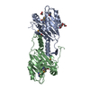
| |||||||||||||||||||||||||||||||||||||||||||||||||||||
|---|---|---|---|---|---|---|---|---|---|---|---|---|---|---|---|---|---|---|---|---|---|---|---|---|---|---|---|---|---|---|---|---|---|---|---|---|---|---|---|---|---|---|---|---|---|---|---|---|---|---|---|---|---|---|
| 1 |
| |||||||||||||||||||||||||||||||||||||||||||||||||||||
| Unit cell |
| |||||||||||||||||||||||||||||||||||||||||||||||||||||
| Noncrystallographic symmetry (NCS) | NCS domain:
NCS domain segments: Component-ID: 1 / Beg label comp-ID: VAL / Refine code: 6
NCS ensembles :
|
-Components
| #1: Protein | Mass: 24090.689 Da / Num. of mol.: 2 Source method: isolated from a genetically manipulated source Source: (gene. exp.) Pseudomonas aeruginosa PAO1 (bacteria) / Species: Pseudomonas aeruginosa / Strain: PAO1, 1C, PRS 101, LMG 12228 / Gene: PA0076 / Plasmid: p11 / Production host: #2: Chemical |   Mass: 59.044 Da / Num. of mol.: 3 / Source method: obtained synthetically / Formula: C2H3O2 Mass: 59.044 Da / Num. of mol.: 3 / Source method: obtained synthetically / Formula: C2H3O2#3: Chemical | ChemComp-PGE / |   Mass: 150.173 Da / Num. of mol.: 1 / Source method: obtained synthetically / Formula: C6H14O4 Mass: 150.173 Da / Num. of mol.: 1 / Source method: obtained synthetically / Formula: C6H14O4#4: Water | ChemComp-HOH / |  Mass: 18.015 Da / Num. of mol.: 120 / Source method: isolated from a natural source / Formula: H2O Mass: 18.015 Da / Num. of mol.: 120 / Source method: isolated from a natural source / Formula: H2O |
|---|
-Experimental details
-Experiment
| Experiment | Method: X-RAY DIFFRACTION / Number of used crystals: 1 |
|---|
- Sample preparation
| Crystal | Density Matthews: 1.99 Å3/Da / Density % sol: 38.13 % |
|---|---|
| Crystal grow | Temperature: 277 K / Method: vapor diffusion, hanging drop / pH: 6.5 Details: 0.1M Sodium cacodylate, 0.2M Calcium acetate, 18% PEG 8000, 0.3M NDSB 256, pH 6.5, VAPOR DIFFUSION, HANGING DROP, temperature 277.0K |
-Data collection
| Diffraction | Mean temperature: 100 K |
|---|---|
| Diffraction source | Source: SYNCHROTRON / Site: APS  / Beamline: 19-ID / Wavelength: 0.9794 Å / Beamline: 19-ID / Wavelength: 0.9794 Å |
| Detector | Type: ADSC QUANTUM 315 / Detector: CCD / Date: Feb 9, 2007 / Details: MIRROR |
| Radiation | Monochromator: SI-111 CHANNEL / Protocol: MAD / Monochromatic (M) / Laue (L): M / Scattering type: x-ray |
| Radiation wavelength | Wavelength: 0.9794 Å / Relative weight: 1 |
| Reflection | Resolution: 2.05→46.32 Å / Num. obs: 23865 / % possible obs: 99.4 % / Observed criterion σ(I): -3 / Redundancy: 9.6 % / Rmerge(I) obs: 0.19 / Rsym value: 0.19 / Net I/σ(I): 17.357 |
| Reflection shell | Resolution: 2.05→2.1 Å / Redundancy: 9.3 % / Rmerge(I) obs: 0.747 / Mean I/σ(I) obs: 3.2 / Rsym value: 0.747 / % possible all: 100 |
- Processing
| Software |
| ||||||||||||||||||||||||||||||||||||||||||||||||||||||||||||||||||||||||||||||||||||||||||||||||||||||||||||||||||||||||||||||||||||||||||||||||||||||||||||||||||||||||||
|---|---|---|---|---|---|---|---|---|---|---|---|---|---|---|---|---|---|---|---|---|---|---|---|---|---|---|---|---|---|---|---|---|---|---|---|---|---|---|---|---|---|---|---|---|---|---|---|---|---|---|---|---|---|---|---|---|---|---|---|---|---|---|---|---|---|---|---|---|---|---|---|---|---|---|---|---|---|---|---|---|---|---|---|---|---|---|---|---|---|---|---|---|---|---|---|---|---|---|---|---|---|---|---|---|---|---|---|---|---|---|---|---|---|---|---|---|---|---|---|---|---|---|---|---|---|---|---|---|---|---|---|---|---|---|---|---|---|---|---|---|---|---|---|---|---|---|---|---|---|---|---|---|---|---|---|---|---|---|---|---|---|---|---|---|---|---|---|---|---|---|---|
| Refinement | Method to determine structure: MAD / Resolution: 2.05→46.32 Å / Cor.coef. Fo:Fc: 0.959 / Cor.coef. Fo:Fc free: 0.941 / SU B: 11.239 / SU ML: 0.151 / Cross valid method: THROUGHOUT / σ(F): 0 / ESU R: 0.233 / ESU R Free: 0.189 / Stereochemistry target values: MAXIMUM LIKELIHOOD / Details: HYDROGENS HAVE BEEN ADDED IN THE RIDING POSITIONS
| ||||||||||||||||||||||||||||||||||||||||||||||||||||||||||||||||||||||||||||||||||||||||||||||||||||||||||||||||||||||||||||||||||||||||||||||||||||||||||||||||||||||||||
| Solvent computation | Ion probe radii: 0.8 Å / Shrinkage radii: 0.8 Å / VDW probe radii: 1.2 Å / Solvent model: MASK | ||||||||||||||||||||||||||||||||||||||||||||||||||||||||||||||||||||||||||||||||||||||||||||||||||||||||||||||||||||||||||||||||||||||||||||||||||||||||||||||||||||||||||
| Displacement parameters | Biso mean: 39.32 Å2
| ||||||||||||||||||||||||||||||||||||||||||||||||||||||||||||||||||||||||||||||||||||||||||||||||||||||||||||||||||||||||||||||||||||||||||||||||||||||||||||||||||||||||||
| Refinement step | Cycle: LAST / Resolution: 2.05→46.32 Å
| ||||||||||||||||||||||||||||||||||||||||||||||||||||||||||||||||||||||||||||||||||||||||||||||||||||||||||||||||||||||||||||||||||||||||||||||||||||||||||||||||||||||||||
| Refine LS restraints |
| ||||||||||||||||||||||||||||||||||||||||||||||||||||||||||||||||||||||||||||||||||||||||||||||||||||||||||||||||||||||||||||||||||||||||||||||||||||||||||||||||||||||||||
| Refine LS restraints NCS | Dom-ID: 1 / Auth asym-ID: A / Refine-ID: X-RAY DIFFRACTION
| ||||||||||||||||||||||||||||||||||||||||||||||||||||||||||||||||||||||||||||||||||||||||||||||||||||||||||||||||||||||||||||||||||||||||||||||||||||||||||||||||||||||||||
| LS refinement shell | Resolution: 2.05→2.1 Å / Total num. of bins used: 20
|
