- PDB-2qiw: Crystal structure of a putative phosphoenolpyruvate phosphonomuta... -
+
Open data
ID or keywords:
Loading...
-
Basic information
Entry
Database: PDB / ID: 2qiw
Title
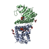
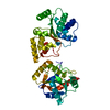
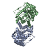
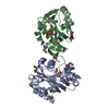
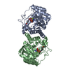
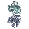
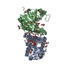
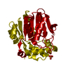
Crystal structure of a putative phosphoenolpyruvate phosphonomutase (ncgl1015, cgl1060) from corynebacterium glutamicum atcc 13032 at 1.80 A resolution
Components
PEP phosphonomutase
Keywords
TRANSFERASE / Structural genomics / Joint Center for Structural Genomics / JCSG / Protein Structure Initiative / PSI-2
Function / homology
Function and homology information
carboxyvinyl-carboxyphosphonate phosphorylmutase / carboxyvinyl-carboxyphosphonate phosphorylmutase activity Similarity search - Function
Phosphoenolpyruvate phosphomutase / Single helix bin / ICL/PEPM domain / Phosphoenolpyruvate-binding domains / Pyruvate kinase-like domain superfamily / Pyruvate/Phosphoenolpyruvate kinase-like domain superfamily / Single alpha-helices involved in coiled-coils or other helix-helix interfaces / TIM Barrel / Alpha-Beta Barrel / Up-down Bundle ...Phosphoenolpyruvate phosphomutase / Single helix bin / ICL/PEPM domain / Phosphoenolpyruvate-binding domains / Pyruvate kinase-like domain superfamily / Pyruvate/Phosphoenolpyruvate kinase-like domain superfamily / Single alpha-helices involved in coiled-coils or other helix-helix interfaces / TIM Barrel / Alpha-Beta Barrel / Up-down Bundle / Mainly Alpha / Alpha Beta Similarity search - Domain/homology
SEQUENCE THE CONSTRUCT WAS EXPRESSED WITH A PURIFICATION TAG MGSDKIHHHHHHENLYFQG. THE TAG WAS ... SEQUENCE THE CONSTRUCT WAS EXPRESSED WITH A PURIFICATION TAG MGSDKIHHHHHHENLYFQG. THE TAG WAS REMOVED WITH TEV PROTEASE LEAVING ONLY A GLYCINE (0) FOLLOWED BY THE TARGET SEQUENCE.
Type: MARMOSAIC 325 mm CCD / Detector: CCD / Date: Jun 21, 2007 / Details: Flat mirror (vertical focusing)
Radiation
Monochromator: Single crystal Si(111) bent (horizontal focusing) Protocol: MAD / Monochromatic (M) / Laue (L): M / Scattering type: x-ray
Radiation wavelength
ID
Wavelength (Å)
Relative weight
1
0.91837
1
2
0.97901
1
3
0.97935
1
Reflection
Resolution: 1.8→29.45 Å / Num. obs: 78759 / % possible obs: 99.6 % / Observed criterion σ(I): -3 / Biso Wilson estimate: 25.945 Å2 / Rmerge(I) obs: 0.056 / Net I/σ(I): 10.6
Reflection shell
Resolution (Å)
Rmerge(I) obs
Mean I/σ(I) obs
Num. measured obs
Num. unique obs
Diffraction-ID
% possible all
1.8-1.86
0.411
2
22993
14091
1
99.4
1.86-1.94
0.318
2.6
26413
16214
1
99.7
1.94-2.03
0.229
3.5
25177
15344
1
99.9
2.03-2.13
0.168
4.9
23210
14131
1
99.7
2.13-2.27
0.127
6.2
26198
15841
1
99.7
2.27-2.44
0.093
8.4
24339
14662
1
99.8
2.44-2.69
0.074
10.2
25670
15366
1
99.7
2.69-3.07
0.047
14.8
24893
14820
1
99.8
3.07-3.87
0.029
22.8
25808
15221
1
99.7
3.87-29.45
0.021
30.5
25985
15029
1
98.5
-
Phasing
Phasing
Method: MAD
-
Processing
Software
Name
Version
Classification
NB
REFMAC
5.2.0019
refinement
PHENIX
refinement
SHELX
phasing
MolProbity
3beta29
modelbuilding
XSCALE
datascaling
PDB_EXTRACT
3
dataextraction
MAR345
CCD
datacollection
XDS
datareduction
SHELXD
phasing
SHARP
phasing
Refinement
Method to determine structure: MAD / Resolution: 1.8→29.45 Å / Cor.coef. Fo:Fc: 0.971 / Cor.coef. Fo:Fc free: 0.96 / SU B: 3.233 / SU ML: 0.051 / TLS residual ADP flag: LIKELY RESIDUAL / Cross valid method: THROUGHOUT / σ(F): 0 / ESU R: 0.074 / ESU R Free: 0.075 Stereochemistry target values: MAXIMUM LIKELIHOOD WITH PHASES Details: 1. HYDROGENS HAVE BEEN ADDED IN THE RIDING POSITIONS. 2. ATOM RECORD CONTAINS RESIDUAL B FACTORS ONLY. 3. A MET-INHIBITION PROTOCOL WAS USED FOR SELENOMETHIONINE INCORPORATION DURING PROTEIN ...Details: 1. HYDROGENS HAVE BEEN ADDED IN THE RIDING POSITIONS. 2. ATOM RECORD CONTAINS RESIDUAL B FACTORS ONLY. 3. A MET-INHIBITION PROTOCOL WAS USED FOR SELENOMETHIONINE INCORPORATION DURING PROTEIN EXPRESSION. THE OCCUPANCY OF THE SE ATOMS IN THE MSE RESIDUES WAS REDUCED TO 0.75 TO ACCOUNT FOR THE REDUCED SCATTERING POWER DUE TO PARTIAL S-MET INCORPORATION. 4. PEG 400 (PG4), HEPES (EPE), SULFATE (SO4), AND GLYCEROL (GOL) ARE PRESENT IN THE CRYSTALLIZATION CRYO BUFFER, AND HAVE BEEN MODELED INTO THE STRUCTURE. 5. AN UNKNOWN LIGAND (UNL) HAS BEEN MODELED INTO THE PUTATIVE ACTIVE SITE ON SUBUNIT A. 6). UNEXPLAINED ELECTRON DENSITIES NEAR HIS 211 IN THE A SUBUNIT, ARG 136 IN THE A SUBUNIT, AND HIS 211 IN THE B SUBUNIT WERE NOT MODELED.
Rfactor
Num. reflection
% reflection
Selection details
Rfree
0.168
3957
5 %
RANDOM
Rwork
0.144
-
-
-
obs
0.145
78697
99.81 %
-
Solvent computation
Ion probe radii: 0.8 Å / Shrinkage radii: 0.8 Å / VDW probe radii: 1.2 Å / Solvent model: BABINET MODEL WITH MASK
In the structure databanks used in Yorodumi, some data are registered as the other names, "COVID-19 virus" and "2019-nCoV". Here are the details of the virus and the list of structure data.
Jan 31, 2019. EMDB accession codes are about to change! (news from PDBe EMDB page)
EMDB accession codes are about to change! (news from PDBe EMDB page)
The allocation of 4 digits for EMDB accession codes will soon come to an end. Whilst these codes will remain in use, new EMDB accession codes will include an additional digit and will expand incrementally as the available range of codes is exhausted. The current 4-digit format prefixed with “EMD-” (i.e. EMD-XXXX) will advance to a 5-digit format (i.e. EMD-XXXXX), and so on. It is currently estimated that the 4-digit codes will be depleted around Spring 2019, at which point the 5-digit format will come into force.
The EM Navigator/Yorodumi systems omit the EMD- prefix.
Related info.:Q: What is EMD? / ID/Accession-code notation in Yorodumi/EM Navigator
Yorodumi is a browser for structure data from EMDB, PDB, SASBDB, etc.
This page is also the successor to EM Navigator detail page, and also detail information page/front-end page for Omokage search.
The word "yorodu" (or yorozu) is an old Japanese word meaning "ten thousand". "mi" (miru) is to see.
Related info.:EMDB / PDB / SASBDB / Comparison of 3 databanks / Yorodumi Search / Aug 31, 2016. New EM Navigator & Yorodumi / Yorodumi Papers / Jmol/JSmol / Function and homology information / Changes in new EM Navigator and Yorodumi
 Movie
Movie Controller

 Yorodumi
 Open data
 Basic information
 Components
 Keywords
 Function and homology information
 Corynebacterium glutamicum ATCC 13032 (bacteria)
 X-RAY DIFFRACTION /
  Authors
 Citation
 Structure visualization
 Downloads & links
 Other downloads

 PDBj
PDBj


 Assembly
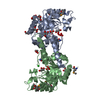


 Mass: 96.063 Da / Num. of mol.: 2 / Source method: obtained synthetically / Formula: SO4
 Mass: 92.094 Da / Num. of mol.: 11 / Source method: obtained synthetically / Formula: C3H8O3
 Mass: 35.453 Da / Num. of mol.: 1 / Source method: obtained synthetically / Formula: Cl
 Mass: 282.331 Da / Num. of mol.: 1 / Source method: obtained synthetically / Formula: C12H26O7 / Comment: precipitant*YM
 Sample preparation
 / Beamline: BL11-1 / Wavelength: 0.91837, 0.97901, 0.97935
 Processing