 Movie
Movie Controller

[English] 日本語
 Yorodumi
- PDB-2pvb: PIKE PARVALBUMIN (PI 4.10) AT LOW TEMPERATURE (100K) AND ATOMIC R... -
+ Open data

- Basic information
| Entry | Database: PDB / ID: 2pvb | ||||||
|---|---|---|---|---|---|---|---|
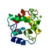
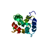
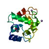
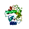
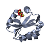
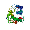
| Title | PIKE PARVALBUMIN (PI 4.10) AT LOW TEMPERATURE (100K) AND ATOMIC RESOLUTION (0.91 A). | ||||||
 Components Components | PROTEIN (PARVALBUMIN) | ||||||
 Keywords Keywords | METAL BINDING PROTEIN / CALCIUM BINDING PROTEIN | ||||||
| Function / homology |  Function and homology information Function and homology information | ||||||
| Biological species |  Esox lucius (northern pike) Esox lucius (northern pike) | ||||||
| Method |  X-RAY DIFFRACTION / SYNCHROTRON / OTHER / Resolution: 0.91 Å X-RAY DIFFRACTION / SYNCHROTRON / OTHER / Resolution: 0.91 Å | ||||||
 Authors Authors | Declercq, J.P. / Evrard, C. | ||||||
 Citation Citation | Journal: Protein Sci. / Year: 1999 Title: Crystal structure of the EF-hand parvalbumin at atomic resolution (0.91 A) and at low temperature (100 K). Evidence for conformational multistates within the hydrophobic core. Authors: Declercq, J.P. / Evrard, C. / Lamzin, V. / Parello, J. #1: Journal: J.Cryst.Growth / Year: 1999Title: A Crystal of a Typical EF-Hand Protein Grown Under Microgravity Diffracts X- Rays Beyond 0.9 A Resolution Authors: Declercq, J.P. / Evrard, C. / Carter, D.C. / Wright, B.S. / Etienne, G. / Parello, J. #2: Journal: Acta Crystallogr.,Sect.D / Year: 1996Title: X-Ray Structure of a New Crystal Form of Pike 4.10 Beta Parvalbumin Authors: Declercq, J.P. / Tinant, B. / Parello, J. #3: Journal: J.Mol.Biol. / Year: 1991Title: Ionic Interactions with Parvalbumins. Crystal Structure Determination of Pike 4.10 Parvalbumin in Four Different Ionic Environments Authors: Declercq, J.P. / Tinant, B. / Parello, J. / Rambaud, J. #4: Journal: J.Mol.Biol. / Year: 1988Title: Crystal Structure Determination and Refinement of Pike 4.10 Parvalbumin (Minor Component from Esox Lucius) Authors: Declercq, J.P. / Tinant, B. / Parello, J. / Etienne, G. / Huber, R. | ||||||
| History |
|
- Structure visualization
| Structure viewer | Molecule: MolmilJmol/JSmol |
|---|
- Downloads & links
-Download
| PDBx/mmCIF format | 2pvb.cif.gz | 66.4 KB | Display | PDBx/mmCIF format |
|---|---|---|---|---|
| PDB format | pdb2pvb.ent.gz | 48.3 KB | Display | PDB format |
| PDBx/mmJSON format | 2pvb.json.gz | Tree view | PDBx/mmJSON format | |
| Others |  Other downloads Other downloads |
-Validation report
| Arichive directory | https://data.pdbj.org/pub/pdb/validation_reports/pv/2pvbftp://data.pdbj.org/pub/pdb/validation_reports/pv/2pvb | HTTPS FTP |
|---|
-Related structure data
| Related structure data |  1pvbS S: Starting model for refinement |
|---|---|
| Similar structure data |


-Links
 PDBj
PDBj
- Assembly
| Deposited unit | 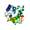
| ||||||||
|---|---|---|---|---|---|---|---|---|---|
| 1 |
| ||||||||
| Unit cell |
|
-Components
| #1: Protein | Mass: 11429.834 Da / Num. of mol.: 1 / Source method: isolated from a natural source / Details: (PIKE PI 4.10) / Source: (natural) Esox lucius (northern pike) / Tissue: MUSCLE / References: UniProt: P02619 | ||||||||||
|---|---|---|---|---|---|---|---|---|---|---|---|
| #2: Chemical |   Mass: 40.078 Da / Num. of mol.: 2 / Source method: obtained synthetically / Formula: Ca Mass: 40.078 Da / Num. of mol.: 2 / Source method: obtained synthetically / Formula: Ca#3: Chemical | ChemComp-NH4 / |   Mass: 18.038 Da / Num. of mol.: 1 / Source method: obtained synthetically / Formula: H4N Mass: 18.038 Da / Num. of mol.: 1 / Source method: obtained synthetically / Formula: H4N#4: Chemical |   Mass: 46.025 Da / Num. of mol.: 2 / Source method: obtained synthetically / Formula: CH2O2 Mass: 46.025 Da / Num. of mol.: 2 / Source method: obtained synthetically / Formula: CH2O2#5: Water | ChemComp-HOH / |  Mass: 18.015 Da / Num. of mol.: 211 / Source method: isolated from a natural source / Formula: H2O Mass: 18.015 Da / Num. of mol.: 211 / Source method: isolated from a natural source / Formula: H2OHas protein modification | Y | Nonpolymer details | ACETYL IS COVALENTLY | |
-Experimental details
-Experiment
| Experiment | Method: X-RAY DIFFRACTION / Number of used crystals: 1 |
|---|
- Sample preparation
| Crystal | Density Matthews: 1.93 Å3/Da / Density % sol: 36.2 % / Description: STRUCTURE PREVIOUSLY SOLVED AT 1.75 A | ||||||||||||||||||||||||||||||
|---|---|---|---|---|---|---|---|---|---|---|---|---|---|---|---|---|---|---|---|---|---|---|---|---|---|---|---|---|---|---|---|
| Crystal grow | Method: vapor diffusion - sitting drop in microgravity / pH: 8 Details: SITTING DROP, MICROGRAVITY CONDITIONS PROTEIN SOLUTION : 27 MG/ML, 0.02% (W/V) NAN3 RESERVOIR : 2.4M AMMONIUM SULFATE 0.03%(W/V) EDTA, 0.02% (W/V) NAN3 TRIS BUFFER (PH 8.0) DROP : 10 MICROL. ...Details: SITTING DROP, MICROGRAVITY CONDITIONS PROTEIN SOLUTION : 27 MG/ML, 0.02% (W/V) NAN3 RESERVOIR : 2.4M AMMONIUM SULFATE 0.03%(W/V) EDTA, 0.02% (W/V) NAN3 TRIS BUFFER (PH 8.0) DROP : 10 MICROL. PROTEIN SOLUTION + 10 MICROL. RESERVOIR, vapor diffusion - sitting drop in microgravity | ||||||||||||||||||||||||||||||
| Crystal grow | *PLUS Method: vapor diffusion, sitting dropDetails: under microgravity, Declercq, J.P., (1999) J.Cryst.Growth, 196, 595. PH range low: 8.25 / PH range high: 7.75 | ||||||||||||||||||||||||||||||
| Components of the solutions | *PLUS
|
-Data collection
| Diffraction | Mean temperature: 100 K |
|---|---|
| Diffraction source | Source: SYNCHROTRON / Site: EMBL/DESY, HAMBURG  / Beamline: X11 / Wavelength: 0.9096 / Beamline: X11 / Wavelength: 0.9096 |
| Detector | Type: MARRESEARCH / Detector: IMAGE PLATE / Date: Jun 1, 1997 / Details: SEGMENTED MIRROR |
| Radiation | Monochromator: BENT SINGLE-CRYSTAL GERMANIUM TRIANGULAR MONOCHROMATOR Protocol: SINGLE WAVELENGTH / Monochromatic (M) / Laue (L): M / Scattering type: x-ray |
| Radiation wavelength | Wavelength: 0.9096 Å / Relative weight: 1 |
| Reflection | Resolution: 0.91→25 Å / Num. obs: 63698 / % possible obs: 99.8 % / Redundancy: 4.9 % / Rsym value: 3.6 / Net I/σ(I): 14.2 |
| Reflection shell | Resolution: 0.91→0.92 Å / Redundancy: 2.8 % / Mean I/σ(I) obs: 3.3 / Rsym value: 12.4 / % possible all: 97.3 |
| Reflection | *PLUS Num. measured all: 481972 / Rmerge(I) obs: 0.036 |
- Processing
| Software |
| |||||||||||||||||||||||||||||||||
|---|---|---|---|---|---|---|---|---|---|---|---|---|---|---|---|---|---|---|---|---|---|---|---|---|---|---|---|---|---|---|---|---|---|---|
| Refinement | Method to determine structure: OTHER Starting model: PDB ENTRY 1PVB Resolution: 0.91→25 Å / Num. parameters: 9883 / Num. restraintsaints: 12405 / Cross valid method: THROUGHOUT / σ(F): 0 / Stereochemistry target values: ENGH AND HUBER
| |||||||||||||||||||||||||||||||||
| Solvent computation | Solvent model: MOEWS & KRETSINGER | |||||||||||||||||||||||||||||||||
| Refine analyze | Num. disordered residues: 16 / Occupancy sum hydrogen: 784 / Occupancy sum non hydrogen: 1025 | |||||||||||||||||||||||||||||||||
| Refinement step | Cycle: LAST / Resolution: 0.91→25 Å
| |||||||||||||||||||||||||||||||||
| Refine LS restraints |
| |||||||||||||||||||||||||||||||||
| Software | *PLUS Name: SHELXL-97 / Classification: refinement | |||||||||||||||||||||||||||||||||
| Refinement | *PLUS σ(F): 0 / % reflection Rfree: 5 % / Rfactor all: 0.11 / Rfactor Rfree: 0.132 / Rfactor Rwork: 0.109 | |||||||||||||||||||||||||||||||||
| Solvent computation | *PLUS | |||||||||||||||||||||||||||||||||
| Displacement parameters | *PLUS | |||||||||||||||||||||||||||||||||
| Refine LS restraints | *PLUS
| |||||||||||||||||||||||||||||||||
| LS refinement shell | *PLUS Highest resolution: 0.91 Å / Lowest resolution: 0.94 Å / Rfactor all: 0.132 |
