 Movie
Movie Controller

[English] 日本語
 Yorodumi
- PDB-4pal: IONIC INTERACTIONS WITH PARVALBUMINS. CRYSTAL STRUCTURE DETERMINA... -
+ Open data

- Basic information
| Entry | Database: PDB / ID: 4pal | ||||||
|---|---|---|---|---|---|---|---|
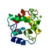
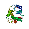
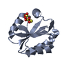
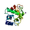
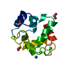
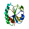
| Title | IONIC INTERACTIONS WITH PARVALBUMINS. CRYSTAL STRUCTURE DETERMINATION OF PIKE 4.10 PARVALBUMIN IN FOUR DIFFERENT IONIC ENVIRONMENTS | ||||||
 Components Components | PARVALBUMIN | ||||||
 Keywords Keywords | CALCIUM BINDING PROTEIN | ||||||
| Function / homology |  Function and homology information Function and homology information | ||||||
| Biological species |  Esox lucius (northern pike) Esox lucius (northern pike) | ||||||
| Method |  X-RAY DIFFRACTION / Resolution: 1.8 Å X-RAY DIFFRACTION / Resolution: 1.8 Å | ||||||
 Authors Authors | Declercq, J.P. / Tinant, B. / Parello, J. / Rambaud, J. | ||||||
 Citation Citation | Journal: J.Mol.Biol. / Year: 1991 Title: Ionic interactions with parvalbumins. Crystal structure determination of pike 4.10 parvalbumin in four different ionic environments. Authors: Declercq, J.P. / Tinant, B. / Parello, J. / Rambaud, J. #1: Journal: J.Mol.Biol. / Year: 1988Title: Crystal Structure Determination and Refinement of Pike 4.10 Parvalbumin (Minor Component from Esox Lucius) Authors: Declercq, J.-P. / Tinant, B. / Parello, J. / Etienne, G. / Huber, R. | ||||||
| History |
|
- Structure visualization
| Structure viewer | Molecule: MolmilJmol/JSmol |
|---|
- Downloads & links
-Download
| PDBx/mmCIF format | 4pal.cif.gz | 35.4 KB | Display | PDBx/mmCIF format |
|---|---|---|---|---|
| PDB format | pdb4pal.ent.gz | 23.8 KB | Display | PDB format |
| PDBx/mmJSON format | 4pal.json.gz | Tree view | PDBx/mmJSON format | |
| Others |  Other downloads Other downloads |
-Validation report
| Arichive directory | https://data.pdbj.org/pub/pdb/validation_reports/pa/4palftp://data.pdbj.org/pub/pdb/validation_reports/pa/4pal | HTTPS FTP |
|---|
-Related structure data


-Links
 PDBj
PDBj- Assembly
| Deposited unit | 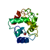
| ||||||||
|---|---|---|---|---|---|---|---|---|---|
| 1 |
| ||||||||
| Unit cell |
|
-Components
| #1: Protein | Mass: 11429.834 Da / Num. of mol.: 1 Source method: isolated from a genetically manipulated source Source: (gene. exp.) Esox lucius (northern pike) / References: UniProt: P02619 | ||||||
|---|---|---|---|---|---|---|---|
| #2: Chemical |   Mass: 24.305 Da / Num. of mol.: 2 / Source method: obtained synthetically / Formula: Mg Mass: 24.305 Da / Num. of mol.: 2 / Source method: obtained synthetically / Formula: Mg#3: Chemical | ChemComp-CA / |   Mass: 40.078 Da / Num. of mol.: 1 / Source method: obtained synthetically / Formula: Ca Mass: 40.078 Da / Num. of mol.: 1 / Source method: obtained synthetically / Formula: Ca#4: Water | ChemComp-HOH / |  Mass: 18.015 Da / Num. of mol.: 88 / Source method: isolated from a natural source / Formula: H2O Mass: 18.015 Da / Num. of mol.: 88 / Source method: isolated from a natural source / Formula: H2OHas protein modification | Y | |
-Experimental details
-Experiment
| Experiment | Method: X-RAY DIFFRACTION |
|---|
- Sample preparation
| Crystal | Density Matthews: 3.16 Å3/Da / Density % sol: 61.08 % |
|---|---|
| Crystal grow | *PLUS Temperature: 20 ℃ / Method: vapor diffusion, hanging drop |
| Components of the solutions | *PLUS Conc.: 3 mM / Common name: sodium azide |
-Data collection
| Reflection | *PLUS Highest resolution: 1.8 Å / Lowest resolution: 9999 Å / Num. obs: 13286 / % possible obs: 89 % / Observed criterion σ(I): 2 / Num. measured all: 64669 / Rmerge(I) obs: 0.089 |
|---|---|
| Reflection shell | *PLUS Highest resolution: 1.8 Å / Lowest resolution: 2.4 Å / % possible obs: 85 % |
- Processing
| Software | Name: EREF / Classification: refinement | ||||||||||||||||||||||||||||||||||||||||||||||||||||||||||||
|---|---|---|---|---|---|---|---|---|---|---|---|---|---|---|---|---|---|---|---|---|---|---|---|---|---|---|---|---|---|---|---|---|---|---|---|---|---|---|---|---|---|---|---|---|---|---|---|---|---|---|---|---|---|---|---|---|---|---|---|---|---|
| Refinement | Rfactor Rwork: 0.18 / Highest resolution: 1.8 Å | ||||||||||||||||||||||||||||||||||||||||||||||||||||||||||||
| Refinement step | Cycle: LAST / Highest resolution: 1.8 Å
| ||||||||||||||||||||||||||||||||||||||||||||||||||||||||||||
| Refine LS restraints |
| ||||||||||||||||||||||||||||||||||||||||||||||||||||||||||||
| Refinement | *PLUS Lowest resolution: 7 Å | ||||||||||||||||||||||||||||||||||||||||||||||||||||||||||||
| Solvent computation | *PLUS | ||||||||||||||||||||||||||||||||||||||||||||||||||||||||||||
| Displacement parameters | *PLUS | ||||||||||||||||||||||||||||||||||||||||||||||||||||||||||||
| Refine LS restraints | *PLUS
| ||||||||||||||||||||||||||||||||||||||||||||||||||||||||||||
| LS refinement shell | *PLUS Highest resolution: 1.8 Å / Lowest resolution: 2.4 Å / Rfactor all: 0.211 |
