 Movie
Movie Controller

[English] 日本語
 Yorodumi
- PDB-2vm2: Crystal structure of barley thioredoxin h isoform 1 crystallized ... -
+ Open data

- Basic information
| Entry | Database: PDB / ID: 2vm2 | ||||||
|---|---|---|---|---|---|---|---|
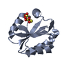
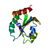
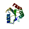
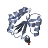
| Title | Crystal structure of barley thioredoxin h isoform 1 crystallized using PEG as precipitant | ||||||
 Components Components | THIOREDOXIN H ISOFORM 1. | ||||||
 Keywords Keywords | OXIDOREDUCTASE / PROTEIN DISULFIDE REDUCTASE / THIOREDOXIN-FOLD | ||||||
| Function / homology |  Function and homology information Function and homology informationplasmodesmata-mediated intercellular transport / oxidoreductase activity, acting on a sulfur group of donors, disulfide as acceptor / protein-disulfide reductase activity / enzyme inhibitor activity / response to cold / response to oxidative stress / cytoplasm Similarity search - Function | ||||||
| Biological species |  | ||||||
| Method |  X-RAY DIFFRACTION / MOLECULAR REPLACEMENT / Resolution: 1.8 Å X-RAY DIFFRACTION / MOLECULAR REPLACEMENT / Resolution: 1.8 Å | ||||||
 Authors Authors | Maeda, K. / Hagglund, P. / Finnie, C. / Svensson, B. / Henriksen, A. | ||||||
 Citation Citation | Journal: Protein Sci. / Year: 2008 Title: Crystal Structures of Barley Thioredoxin H Isoforms Hvtrxh1 and Hvtrxh2 Reveal Features Involved in Protein Recognition and Possibly in Discriminating the Isoform Specificity. Authors: Maeda, K. / Hagglund, P. / Finnie, C. / Svensson, B. / Henriksen, A. | ||||||
| History |
|
- Structure visualization
| Structure viewer | Molecule: MolmilJmol/JSmol |
|---|
- Downloads & links
-Download
| PDBx/mmCIF format | 2vm2.cif.gz | 106.1 KB | Display | PDBx/mmCIF format |
|---|---|---|---|---|
| PDB format | pdb2vm2.ent.gz | 83.1 KB | Display | PDB format |
| PDBx/mmJSON format | 2vm2.json.gz | Tree view | PDBx/mmJSON format | |
| Others |  Other downloads Other downloads |
-Validation report
| Arichive directory | https://data.pdbj.org/pub/pdb/validation_reports/vm/2vm2ftp://data.pdbj.org/pub/pdb/validation_reports/vm/2vm2 | HTTPS FTP |
|---|
-Related structure data
| Related structure data |  2vltC 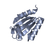 2vluC  2vlvC  2vm1C 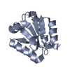 1ep7S C: citing same article ( S: Starting model for refinement |
|---|---|
| Similar structure data |


-Links
 PDBj
PDBj

- Assembly
| Deposited unit | 
| ||||||||
|---|---|---|---|---|---|---|---|---|---|
| 1 | 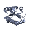
| ||||||||
| 2 | 
| ||||||||
| 3 | 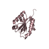
| ||||||||
| 4 | 
| ||||||||
| Unit cell |
|
-Components
| #1: Protein | Mass: 12769.681 Da / Num. of mol.: 4 Source method: isolated from a genetically manipulated source Source: (gene. exp.) Variant: BARKE / Plasmid: PET11A / Production host:  References: UniProt: Q7XZK3, thioredoxin-disulfide reductase (NADPH) #2: Water | ChemComp-HOH / |  Mass: 18.015 Da / Num. of mol.: 544 / Source method: isolated from a natural source / Formula: H2O Mass: 18.015 Da / Num. of mol.: 544 / Source method: isolated from a natural source / Formula: H2OHas protein modification | Y | |
|---|
-Experimental details
-Experiment
| Experiment | Method: X-RAY DIFFRACTION |
|---|
- Sample preparation
| Crystal | Density Matthews: 2.27 Å3/Da / Density % sol: 45.41 % / Description: NONE |
|---|
-Data collection
| Diffraction | Mean temperature: 100 K |
|---|---|
| Diffraction source | Source: ROTATING ANODE / Type: RIGAKU RUH3R / Wavelength: 1.5418 |
| Radiation | Protocol: SINGLE WAVELENGTH / Monochromatic (M) / Laue (L): M / Scattering type: x-ray |
| Radiation wavelength | Wavelength: 1.5418 Å / Relative weight: 1 |
| Reflection | Resolution: 1.8→20.6 Å / Num. obs: 46412 / % possible obs: 99.3 % / Observed criterion σ(I): 0 / Redundancy: 2.9 % / Rmerge(I) obs: 0.07 / Net I/σ(I): 6.5 |
| Reflection shell | Resolution: 1.8→1.9 Å / Redundancy: 2.8 % / Rmerge(I) obs: 0.35 / Mean I/σ(I) obs: 2.1 / % possible all: 99.8 |
- Processing
| Software | Name: REFMAC / Version: 5.2.0005 / Classification: refinement | ||||||||||||||||||||||||||||||||||||||||||||||||||||||||||||||||||||||||||||||||||||||||||||||||||||||||||||||||||||||||||||||||||||||||||||||||||||||||||||||||||||||||||||||||||||||
|---|---|---|---|---|---|---|---|---|---|---|---|---|---|---|---|---|---|---|---|---|---|---|---|---|---|---|---|---|---|---|---|---|---|---|---|---|---|---|---|---|---|---|---|---|---|---|---|---|---|---|---|---|---|---|---|---|---|---|---|---|---|---|---|---|---|---|---|---|---|---|---|---|---|---|---|---|---|---|---|---|---|---|---|---|---|---|---|---|---|---|---|---|---|---|---|---|---|---|---|---|---|---|---|---|---|---|---|---|---|---|---|---|---|---|---|---|---|---|---|---|---|---|---|---|---|---|---|---|---|---|---|---|---|---|---|---|---|---|---|---|---|---|---|---|---|---|---|---|---|---|---|---|---|---|---|---|---|---|---|---|---|---|---|---|---|---|---|---|---|---|---|---|---|---|---|---|---|---|---|---|---|---|---|
| Refinement | Method to determine structure: MOLECULAR REPLACEMENT Starting model: PDB ENTRY 1EP7 Resolution: 1.8→20.36 Å / Cor.coef. Fo:Fc: 0.948 / Cor.coef. Fo:Fc free: 0.921 / SU B: 5.224 / SU ML: 0.088 / TLS residual ADP flag: LIKELY RESIDUAL / Cross valid method: THROUGHOUT / ESU R: 0.134 / ESU R Free: 0.137 / Stereochemistry target values: MAXIMUM LIKELIHOOD / Details: HYDROGENS HAVE BEEN ADDED IN THE RIDING POSITIONS.
| ||||||||||||||||||||||||||||||||||||||||||||||||||||||||||||||||||||||||||||||||||||||||||||||||||||||||||||||||||||||||||||||||||||||||||||||||||||||||||||||||||||||||||||||||||||||
| Solvent computation | Ion probe radii: 0.8 Å / Shrinkage radii: 0.8 Å / VDW probe radii: 1.2 Å / Solvent model: MASK | ||||||||||||||||||||||||||||||||||||||||||||||||||||||||||||||||||||||||||||||||||||||||||||||||||||||||||||||||||||||||||||||||||||||||||||||||||||||||||||||||||||||||||||||||||||||
| Displacement parameters | Biso mean: 24.67 Å2
| ||||||||||||||||||||||||||||||||||||||||||||||||||||||||||||||||||||||||||||||||||||||||||||||||||||||||||||||||||||||||||||||||||||||||||||||||||||||||||||||||||||||||||||||||||||||
| Refinement step | Cycle: LAST / Resolution: 1.8→20.36 Å
| ||||||||||||||||||||||||||||||||||||||||||||||||||||||||||||||||||||||||||||||||||||||||||||||||||||||||||||||||||||||||||||||||||||||||||||||||||||||||||||||||||||||||||||||||||||||
| Refine LS restraints |
|