 Movie
Movie Controller


+ Open data

- Basic information
| Entry | Database: PDB / ID: 2hxk | ||||||
|---|---|---|---|---|---|---|---|
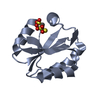
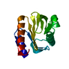
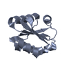
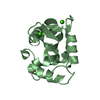
| Title | Crystal structure of S-nitroso thioredoxin | ||||||
 Components Components | Thioredoxin | ||||||
 Keywords Keywords | OXIDOREDUCTASE / S-nitrosation / S-nitrosocysteine | ||||||
| Function / homology |  Function and homology information Function and homology informationProtein repair / cellular detoxification of hydrogen peroxide / protein-disulfide reductase [NAD(P)H] activity / Interconversion of nucleotide di- and triphosphates / thioredoxin-disulfide reductase (NADPH) activity / negative regulation of protein export from nucleus / Regulation of FOXO transcriptional activity by acetylation / positive regulation of DNA binding / NFE2L2 regulating anti-oxidant/detoxification enzymes / response to nitric oxide ...Protein repair / cellular detoxification of hydrogen peroxide / protein-disulfide reductase [NAD(P)H] activity / Interconversion of nucleotide di- and triphosphates / thioredoxin-disulfide reductase (NADPH) activity / negative regulation of protein export from nucleus / Regulation of FOXO transcriptional activity by acetylation / positive regulation of DNA binding / NFE2L2 regulating anti-oxidant/detoxification enzymes / response to nitric oxide / Detoxification of Reactive Oxygen Species / protein-disulfide reductase activity / The NLRP3 inflammasome / Purinergic signaling in leishmaniasis infection / cell redox homeostasis / TP53 Regulates Metabolic Genes / response to radiation / Oxidative Stress Induced Senescence / positive regulation of phosphatidylinositol 3-kinase/protein kinase B signal transduction / negative regulation of transcription by RNA polymerase II / protein homodimerization activity / RNA binding / extracellular exosome / extracellular region / nucleoplasm / nucleus / cytosol / cytoplasm Similarity search - Function | ||||||
| Biological species |  Homo sapiens (human) Homo sapiens (human) | ||||||
| Method |  X-RAY DIFFRACTION / MOLECULAR REPLACEMENT / Resolution: 1.65 Å X-RAY DIFFRACTION / MOLECULAR REPLACEMENT / Resolution: 1.65 Å | ||||||
 Authors Authors | Weichsel, A. / Montfort, W.R. | ||||||
 Citation Citation | Journal: Biochemistry / Year: 2007 Title: Buried s-nitrosocysteine revealed in crystal structures of human thioredoxin. Authors: Weichsel, A. / Brailey, J.L. / Montfort, W.R. #1: Journal: Structure / Year: 1996Title: Crystal structures of reduced, oxidized, and mutated human thioredoxins: evidence for a regulatory homodimer Authors: Weichsel, A. / Gasdaska, J.R. / Powis, G. / Montfort, W.R. | ||||||
| History |
|
- Structure visualization
| Structure viewer | Molecule: MolmilJmol/JSmol |
|---|
- Downloads & links
-Download
| PDBx/mmCIF format | 2hxk.cif.gz | 82 KB | Display | PDBx/mmCIF format |
|---|---|---|---|---|
| PDB format | pdb2hxk.ent.gz | 63.8 KB | Display | PDB format |
| PDBx/mmJSON format | 2hxk.json.gz | Tree view | PDBx/mmJSON format | |
| Others |  Other downloads Other downloads |
-Validation report
| Arichive directory | https://data.pdbj.org/pub/pdb/validation_reports/hx/2hxkftp://data.pdbj.org/pub/pdb/validation_reports/hx/2hxk | HTTPS FTP |
|---|
-Related structure data
| Related structure data |  2hshC  2ifqC  2iiyC  1ertS S: Starting model for refinement C: citing same article ( |
|---|---|
| Similar structure data |


-Links
 PDBj
PDBj


- Assembly
| Deposited unit | 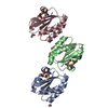
| ||||||||
|---|---|---|---|---|---|---|---|---|---|
| 1 | 
| ||||||||
| 2 | 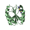
| ||||||||
| 3 | 
| ||||||||
| Unit cell |
|
-Components
| #1: Protein | Mass: 11808.474 Da / Num. of mol.: 3 Source method: isolated from a genetically manipulated source Source: (gene. exp.) Homo sapiens (human) / Gene: TXN / Plasmid: pet-3a / Production host:  #2: Chemical |   Mass: 46.068 Da / Num. of mol.: 3 / Source method: obtained synthetically / Formula: C2H6O Mass: 46.068 Da / Num. of mol.: 3 / Source method: obtained synthetically / Formula: C2H6O#3: Water | ChemComp-HOH / |  Mass: 18.015 Da / Num. of mol.: 139 / Source method: isolated from a natural source / Formula: H2O Mass: 18.015 Da / Num. of mol.: 139 / Source method: isolated from a natural source / Formula: H2OHas protein modification | Y | |
|---|
-Experimental details
-Experiment
| Experiment | Method: X-RAY DIFFRACTION / Number of used crystals: 1 |
|---|
- Sample preparation
| Crystal | Density Matthews: 1.66 Å3/Da / Density % sol: 25.13 % |
|---|---|
| Crystal grow | Temperature: 298 K / Method: vapor diffusion, hanging drop / pH: 7 Details: 32% PEG 1500, 50 mM sodium phosphate, ethanol, pH 7.0, VAPOR DIFFUSION, HANGING DROP, temperature 298K |
-Data collection
| Diffraction | Mean temperature: 100 K |
|---|---|
| Diffraction source | Source: ROTATING ANODE / Type: RIGAKU RU300 / Wavelength: 1.5418 Å |
| Detector | Type: RIGAKU RAXIS IV / Detector: IMAGE PLATE / Date: Mar 8, 2006 / Details: osmic mirrors |
| Radiation | Monochromator: osmic mirrors / Protocol: SINGLE WAVELENGTH / Monochromatic (M) / Laue (L): M / Scattering type: x-ray |
| Radiation wavelength | Wavelength: 1.5418 Å / Relative weight: 1 |
| Reflection | Resolution: 1.65→24.2 Å / Num. all: 29229 / Num. obs: 29229 / % possible obs: 99 % / Observed criterion σ(F): 0 / Observed criterion σ(I): 0 / Redundancy: 2.7 % / Biso Wilson estimate: 26 Å2 / Rmerge(I) obs: 0.04 / Net I/σ(I): 16.1 |
| Reflection shell | Resolution: 1.65→1.71 Å / Redundancy: 2.2 % / Rmerge(I) obs: 0.14 / Mean I/σ(I) obs: 3.6 / Num. unique all: 3122 / % possible all: 99.9 |
- Processing
| Software |
| ||||||||||||||||||||||||||||||||||||||||||||||||||||||||||||||||||||||||||||||||||||||||||||||||||||||||||||||||||||||||
|---|---|---|---|---|---|---|---|---|---|---|---|---|---|---|---|---|---|---|---|---|---|---|---|---|---|---|---|---|---|---|---|---|---|---|---|---|---|---|---|---|---|---|---|---|---|---|---|---|---|---|---|---|---|---|---|---|---|---|---|---|---|---|---|---|---|---|---|---|---|---|---|---|---|---|---|---|---|---|---|---|---|---|---|---|---|---|---|---|---|---|---|---|---|---|---|---|---|---|---|---|---|---|---|---|---|---|---|---|---|---|---|---|---|---|---|---|---|---|---|---|---|
| Refinement | Method to determine structure: MOLECULAR REPLACEMENT Starting model: PDB ENTRY 1ERT Resolution: 1.65→24.2 Å / Cor.coef. Fo:Fc: 0.948 / Cor.coef. Fo:Fc free: 0.93 / SU B: 2.223 / SU ML: 0.079 / Isotropic thermal model: isotropic / Cross valid method: THROUGHOUT / σ(F): 0 / σ(I): 0 / ESU R: 0.133 / ESU R Free: 0.127 / Stereochemistry target values: MAXIMUM LIKELIHOOD / Details: HYDROGENS HAVE BEEN ADDED IN THE RIDING POSITIONS
| ||||||||||||||||||||||||||||||||||||||||||||||||||||||||||||||||||||||||||||||||||||||||||||||||||||||||||||||||||||||||
| Solvent computation | Ion probe radii: 0.8 Å / Shrinkage radii: 0.8 Å / VDW probe radii: 1.4 Å / Solvent model: MASK | ||||||||||||||||||||||||||||||||||||||||||||||||||||||||||||||||||||||||||||||||||||||||||||||||||||||||||||||||||||||||
| Displacement parameters | Biso mean: 13.156 Å2
| ||||||||||||||||||||||||||||||||||||||||||||||||||||||||||||||||||||||||||||||||||||||||||||||||||||||||||||||||||||||||
| Refine analyze | Luzzati coordinate error obs: 0.189 Å | ||||||||||||||||||||||||||||||||||||||||||||||||||||||||||||||||||||||||||||||||||||||||||||||||||||||||||||||||||||||||
| Refinement step | Cycle: LAST / Resolution: 1.65→24.2 Å
| ||||||||||||||||||||||||||||||||||||||||||||||||||||||||||||||||||||||||||||||||||||||||||||||||||||||||||||||||||||||||
| Refine LS restraints |
| ||||||||||||||||||||||||||||||||||||||||||||||||||||||||||||||||||||||||||||||||||||||||||||||||||||||||||||||||||||||||
| LS refinement shell | Resolution: 1.65→1.693 Å / Total num. of bins used: 20
|
