 Movie
Movie Controller

[English] 日本語
 Yorodumi
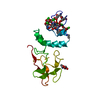
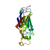
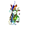
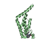
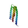
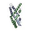
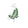
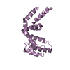
- PDB-2pf2: THE CA+2 ION AND MEMBRANE BINDING STRUCTURE OF THE GLA DOMAIN OF ... -
+ Open data

- Basic information
| Entry | Database: PDB / ID: 2pf2 | ||||||
|---|---|---|---|---|---|---|---|
| Title | THE CA+2 ION AND MEMBRANE BINDING STRUCTURE OF THE GLA DOMAIN OF CA-PROTHROMBIN FRAGMENT 1 | ||||||
 Components Components | PROTHROMBIN FRAGMENT 1 | ||||||
 Keywords Keywords | HYDROLASE(SERINE PROTEASE) | ||||||
| Function / homology |  Function and homology information Function and homology informationfibrinogen binding / thrombin / positive regulation of blood coagulation / protein polymerization / acute-phase response / platelet activation / serine-type endopeptidase activity / calcium ion binding / proteolysis / : Similarity search - Function | ||||||
| Biological species |  | ||||||
| Method |  X-RAY DIFFRACTION / Resolution: 2.2 Å X-RAY DIFFRACTION / Resolution: 2.2 Å | ||||||
 Authors Authors | Soriano-Garcia, M. / Padmanabhan, K. / De vos, A.M. / Tulinsky, A. | ||||||
 Citation Citation | Journal: Biochemistry / Year: 1992 Title: The Ca2+ ion and membrane binding structure of the Gla domain of Ca-prothrombin fragment 1. Authors: Soriano-Garcia, M. / Padmanabhan, K. / de Vos, A.M. / Tulinsky, A. #1: Journal: J.Mol.Biol. / Year: 1991Title: Structure of Bovine Prothrombin Fragment 1 Refined at 2.25 Angstroms Resolution Authors: Seshadri, T.P. / Tulinsky, A. / Skrzypczak-Jankun, E. / Park, C.H. #2: Journal: Biochemistry / Year: 1989Title: Structure of Ca2+ Prothrombin Fragment 1 Including the Conformation of the Gla Domain Authors: Soriano-Garcia, M. / Park, C.H. / Tulinsky, A. / Ravichandran, K.G. / Skrzypczak-Jankun, E. | ||||||
| History |
|
- Structure visualization
| Structure viewer | Molecule: MolmilJmol/JSmol |
|---|
- Downloads & links
-Download
| PDBx/mmCIF format | 2pf2.cif.gz | 50.1 KB | Display | PDBx/mmCIF format |
|---|---|---|---|---|
| PDB format | pdb2pf2.ent.gz | 34.7 KB | Display | PDB format |
| PDBx/mmJSON format | 2pf2.json.gz | Tree view | PDBx/mmJSON format | |
| Others |  Other downloads Other downloads |
-Validation report
| Arichive directory | https://data.pdbj.org/pub/pdb/validation_reports/pf/2pf2ftp://data.pdbj.org/pub/pdb/validation_reports/pf/2pf2 | HTTPS FTP |
|---|
-Related structure data
| Similar structure data |
|---|


-Links
 PDBj
PDBj


- Assembly
| Deposited unit | 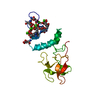
| ||||||||
|---|---|---|---|---|---|---|---|---|---|
| 1 |
| ||||||||
| Unit cell |
| ||||||||
| Atom site foot note | 1: RESIDUES PRO 54 AND PRO 95 ARE CIS PROLINES. |
-Components
| #1: Protein | Mass: 18024.672 Da / Num. of mol.: 1 Source method: isolated from a genetically manipulated source Source: (gene. exp.) | ||||
|---|---|---|---|---|---|
| #2: Chemical | ChemComp-CA /   Mass: 40.078 Da / Num. of mol.: 7 / Source method: obtained synthetically / Formula: Ca Mass: 40.078 Da / Num. of mol.: 7 / Source method: obtained synthetically / Formula: Ca#3: Water | ChemComp-HOH / |  Mass: 18.015 Da / Num. of mol.: 144 / Source method: isolated from a natural source / Formula: H2O Mass: 18.015 Da / Num. of mol.: 144 / Source method: isolated from a natural source / Formula: H2OHas protein modification | Y | |
-Experimental details
-Experiment
| Experiment | Method: X-RAY DIFFRACTION |
|---|
- Sample preparation
| Crystal | Density Matthews: 3.81 Å3/Da / Density % sol: 67.75 % | ||||||||||||||||||||||||||||||||||||||||||
|---|---|---|---|---|---|---|---|---|---|---|---|---|---|---|---|---|---|---|---|---|---|---|---|---|---|---|---|---|---|---|---|---|---|---|---|---|---|---|---|---|---|---|---|
| Crystal grow | *PLUS pH: 6.5 / Method: vapor diffusion, hanging dropDetails: taken from Soriano-Garacia, M. et al (1989), Biochemistry, 28, 6805-6810. | ||||||||||||||||||||||||||||||||||||||||||
| Components of the solutions | *PLUS
|
-Data collection
| Radiation | Scattering type: x-ray |
|---|---|
| Radiation wavelength | Relative weight: 1 |
| Reflection | *PLUS Highest resolution: 2.2 Å / Num. all: 12662 / Num. obs: 8141 / % possible obs: 64 % |
- Processing
| Software |
| ||||||||||||||||||||||||||||||||||||||||||||||||||||||||||||||||||||||||||||||||||||
|---|---|---|---|---|---|---|---|---|---|---|---|---|---|---|---|---|---|---|---|---|---|---|---|---|---|---|---|---|---|---|---|---|---|---|---|---|---|---|---|---|---|---|---|---|---|---|---|---|---|---|---|---|---|---|---|---|---|---|---|---|---|---|---|---|---|---|---|---|---|---|---|---|---|---|---|---|---|---|---|---|---|---|---|---|---|
| Refinement | Resolution: 2.2→7 Å / Rfactor obs: 0.171 / σ(F): 1 | ||||||||||||||||||||||||||||||||||||||||||||||||||||||||||||||||||||||||||||||||||||
| Refinement step | Cycle: LAST / Resolution: 2.2→7 Å
| ||||||||||||||||||||||||||||||||||||||||||||||||||||||||||||||||||||||||||||||||||||
| Refine LS restraints |
| ||||||||||||||||||||||||||||||||||||||||||||||||||||||||||||||||||||||||||||||||||||
| Software | *PLUS Name: PROLSQ / Classification: refinement | ||||||||||||||||||||||||||||||||||||||||||||||||||||||||||||||||||||||||||||||||||||
| Refinement | *PLUS Highest resolution: 2.2 Å / Lowest resolution: 7 Å / Num. reflection obs: 1024 / σ(F): 1 / Rfactor obs: 0.171 | ||||||||||||||||||||||||||||||||||||||||||||||||||||||||||||||||||||||||||||||||||||
| Solvent computation | *PLUS | ||||||||||||||||||||||||||||||||||||||||||||||||||||||||||||||||||||||||||||||||||||
| Displacement parameters | *PLUS |
