 Movie
Movie Controller

[English] 日本語
 Yorodumi
- PDB-2p8u: Crystal structure of human 3-hydroxy-3-methylglutaryl CoA synthase I -
+ Open data

- Basic information
| Entry | Database: PDB / ID: 2p8u | ||||||
|---|---|---|---|---|---|---|---|
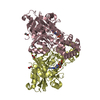
| Title | Crystal structure of human 3-hydroxy-3-methylglutaryl CoA synthase I | ||||||
 Components Components | Hydroxymethylglutaryl-CoA synthase, cytoplasmic | ||||||
 Keywords Keywords | TRANSFERASE / hydromethylglutaryl CoA / mevalonate pathway / Structural Genomics / Structural Genomics Consortium / SGC | ||||||
| Function / homology |  Function and homology information Function and homology informationhydroxymethylglutaryl-CoA synthase / hydroxymethylglutaryl-CoA synthase activity / isopentenyl diphosphate biosynthetic process, mevalonate pathway / Lanosterol biosynthesis / zymosterol biosynthetic process / : / : / farnesyl diphosphate biosynthetic process, mevalonate pathway / geranylgeranyl diphosphate biosynthetic process / acetyl-CoA metabolic process ...hydroxymethylglutaryl-CoA synthase / hydroxymethylglutaryl-CoA synthase activity / isopentenyl diphosphate biosynthetic process, mevalonate pathway / Lanosterol biosynthesis / zymosterol biosynthetic process / : / : / farnesyl diphosphate biosynthetic process, mevalonate pathway / geranylgeranyl diphosphate biosynthetic process / acetyl-CoA metabolic process / cholesterol biosynthetic process / Activation of gene expression by SREBF (SREBP) / lipid metabolic process / PPARA activates gene expression / protein homodimerization activity / nucleoplasm / plasma membrane / cytosol / cytoplasm Similarity search - Function | ||||||
| Biological species |  Homo sapiens (human) Homo sapiens (human) | ||||||
| Method |  X-RAY DIFFRACTION / SYNCHROTRON / MOLECULAR REPLACEMENT / Resolution: 2 Å X-RAY DIFFRACTION / SYNCHROTRON / MOLECULAR REPLACEMENT / Resolution: 2 Å | ||||||
 Authors Authors | Turnbull, A. / Shafqat, N. / Salah, E. / Niesen, F.H. / Burgess, N. / Bunkoczi, G. / Debreczeni, J. / Pike, A.C.W. / Umeano, C. / Gorrec, F. ...Turnbull, A. / Shafqat, N. / Salah, E. / Niesen, F.H. / Burgess, N. / Bunkoczi, G. / Debreczeni, J. / Pike, A.C.W. / Umeano, C. / Gorrec, F. / von Delft, F. / Weigelt, J. / Arrowsmith, C.H. / Sundstrom, M. / Edwards, A. / Oppermann, U. / Structural Genomics Consortium (SGC) | ||||||
 Citation Citation | Journal: J.Mol.Biol. / Year: 2010 Title: Crystal structures of human HMG-CoA synthase isoforms provide insights into inherited ketogenesis disorders and inhibitor design. Authors: Shafqat, N. / Turnbull, A. / Zschocke, J. / Oppermann, U. / Yue, W.W. | ||||||
| History |
|
- Structure visualization
| Structure viewer | Molecule: MolmilJmol/JSmol |
|---|
- Downloads & links
-Download
| PDBx/mmCIF format | 2p8u.cif.gz | 210.5 KB | Display | PDBx/mmCIF format |
|---|---|---|---|---|
| PDB format | pdb2p8u.ent.gz | 165.6 KB | Display | PDB format |
| PDBx/mmJSON format | 2p8u.json.gz | Tree view | PDBx/mmJSON format | |
| Others |  Other downloads Other downloads |
-Validation report
| Arichive directory | https://data.pdbj.org/pub/pdb/validation_reports/p8/2p8uftp://data.pdbj.org/pub/pdb/validation_reports/p8/2p8u | HTTPS FTP |
|---|
-Related structure data
| Related structure data | 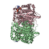 2wyaC 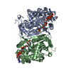 1xplS  2f82S 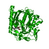 2f9aS 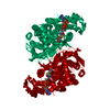 2fa0S  2fa3S S: Starting model for refinement C: citing same article ( |
|---|---|
| Similar structure data |


-Links
 PDBj
PDBj


- Assembly
| Deposited unit | 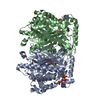
| |||||||||||||||||||||||||||||||||||||||||||||||||||||||||||||||||||||||||||||||||||||||||||||||||||||||||||||||||||||||||||||||||||||||||||||||||||||||||||||||||||||||||||||||||||||||||||||||||||||
|---|---|---|---|---|---|---|---|---|---|---|---|---|---|---|---|---|---|---|---|---|---|---|---|---|---|---|---|---|---|---|---|---|---|---|---|---|---|---|---|---|---|---|---|---|---|---|---|---|---|---|---|---|---|---|---|---|---|---|---|---|---|---|---|---|---|---|---|---|---|---|---|---|---|---|---|---|---|---|---|---|---|---|---|---|---|---|---|---|---|---|---|---|---|---|---|---|---|---|---|---|---|---|---|---|---|---|---|---|---|---|---|---|---|---|---|---|---|---|---|---|---|---|---|---|---|---|---|---|---|---|---|---|---|---|---|---|---|---|---|---|---|---|---|---|---|---|---|---|---|---|---|---|---|---|---|---|---|---|---|---|---|---|---|---|---|---|---|---|---|---|---|---|---|---|---|---|---|---|---|---|---|---|---|---|---|---|---|---|---|---|---|---|---|---|---|---|---|---|
| 1 |
| |||||||||||||||||||||||||||||||||||||||||||||||||||||||||||||||||||||||||||||||||||||||||||||||||||||||||||||||||||||||||||||||||||||||||||||||||||||||||||||||||||||||||||||||||||||||||||||||||||||
| Unit cell |
| |||||||||||||||||||||||||||||||||||||||||||||||||||||||||||||||||||||||||||||||||||||||||||||||||||||||||||||||||||||||||||||||||||||||||||||||||||||||||||||||||||||||||||||||||||||||||||||||||||||
| Noncrystallographic symmetry (NCS) | NCS domain:
NCS domain segments: Ens-ID: 1
|