 Movie
Movie Controller

[English] 日本語
 Yorodumi
- PDB-1xpl: Crystal Structure of Staphylococcus aureus HMG-COA Synthase with ... -
+ Open data

- Basic information
| Entry | Database: PDB / ID: 1xpl | ||||||
|---|---|---|---|---|---|---|---|
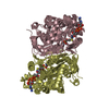
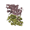
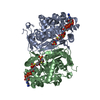
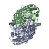
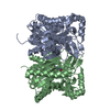
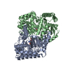
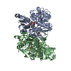
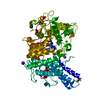
| Title | Crystal Structure of Staphylococcus aureus HMG-COA Synthase with Acetoacetyl-COA and Acetylated Cysteine | ||||||
 Components Components | 3-hydroxy-3-methylglutaryl CoA synthase | ||||||
 Keywords Keywords | TRANSFERASE / HMG-COA Synthase / HMGS / Coenzyme A / Thiolase Fold / Condensing Enzyme / Cholesterol Biosynthesis | ||||||
| Function / homology |  Function and homology information Function and homology informationfarnesyl diphosphate biosynthetic process, mevalonate pathway / hydroxymethylglutaryl-CoA synthase activity / acetyl-CoA metabolic process Similarity search - Function | ||||||
| Biological species |   Staphylococcus aureus subsp. aureus (bacteria) Staphylococcus aureus subsp. aureus (bacteria) | ||||||
| Method |  X-RAY DIFFRACTION / SYNCHROTRON / MOLECULAR REPLACEMENT / Resolution: 2 Å X-RAY DIFFRACTION / SYNCHROTRON / MOLECULAR REPLACEMENT / Resolution: 2 Å | ||||||
 Authors Authors | Theisen, M.J. / Misra, I. / Saadat, D. / Campobasso, N. / Miziorko, H.M. / Harrison, D.H.T. | ||||||
 Citation Citation | Journal: Proc.Natl.Acad.Sci.USA / Year: 2004 Title: 3-hydroxy-3-methylglutaryl-CoA synthase intermediate complex observed in "real-time" Authors: Theisen, M.J. / Misra, I. / Saadat, D. / Campobasso, N. / Miziorko, H.M. / Harrison, D.H.T. | ||||||
| History |
| ||||||
| Remark 999 | SEQUENCE RESULTS ARE FROM DAY FOURTEEN OF CRYSTALLIZATION. IN THE COORDINATE RECORDS SCY/CYS 111 ...SEQUENCE RESULTS ARE FROM DAY FOURTEEN OF CRYSTALLIZATION. IN THE COORDINATE RECORDS SCY/CYS 111 ISOFORMS ARE MODELED AS ALTERNATE CONFORMERS. BECAUSE OF THE FORMAT RESTRICTIONS ONLY SCY 111 ISOFORM IS REPRESENTED IN THE SEQUENCE RECORDS. |
- Structure visualization
| Structure viewer | Molecule: MolmilJmol/JSmol |
|---|
- Downloads & links
-Download
| PDBx/mmCIF format | 1xpl.cif.gz | 320.7 KB | Display | PDBx/mmCIF format |
|---|---|---|---|---|
| PDB format | pdb1xpl.ent.gz | 259.1 KB | Display | PDB format |
| PDBx/mmJSON format | 1xpl.json.gz | Tree view | PDBx/mmJSON format | |
| Others |  Other downloads Other downloads |
-Validation report
| Arichive directory | https://data.pdbj.org/pub/pdb/validation_reports/xp/1xplftp://data.pdbj.org/pub/pdb/validation_reports/xp/1xpl | HTTPS FTP |
|---|
-Related structure data
| Related structure data |  1xpkSC  1xpmC S: Starting model for refinement C: citing same article ( |
|---|---|
| Similar structure data |


-Links
 PDBj
PDBj

- Assembly
| Deposited unit | 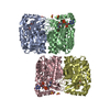
| ||||||||
|---|---|---|---|---|---|---|---|---|---|
| 1 | 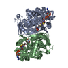
| ||||||||
| 2 | 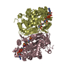
| ||||||||
| Unit cell |
| ||||||||
| Details | The asymmetric unit contains two dimers (A/B and C/D); the dimer is the biological unit. |
-Components
| #1: Protein | Mass: 44335.480 Da / Num. of mol.: 4 Source method: isolated from a genetically manipulated source Source: (gene. exp.) Staphylococcus aureus subsp. aureus (bacteria)Species: Staphylococcus aureus / Strain: subsp. aureus / Gene: mvaS / Plasmid: pET23d / Species (production host): Escherichia coli / Production host: References: UniProt: Q79ZY6, UniProt: A0A0H3K1U2*PLUS, hydroxymethylglutaryl-CoA synthase #2: Chemical | ChemComp-SO4 /   Mass: 96.063 Da / Num. of mol.: 8 / Source method: obtained synthetically / Formula: SO4 Mass: 96.063 Da / Num. of mol.: 8 / Source method: obtained synthetically / Formula: SO4#3: Chemical | ChemComp-CAA /   Mass: 851.607 Da / Num. of mol.: 4 / Source method: obtained synthetically / Formula: C25H40N7O18P3S Mass: 851.607 Da / Num. of mol.: 4 / Source method: obtained synthetically / Formula: C25H40N7O18P3S#4: Water | ChemComp-HOH / |  Mass: 18.015 Da / Num. of mol.: 455 / Source method: isolated from a natural source / Formula: H2O Mass: 18.015 Da / Num. of mol.: 455 / Source method: isolated from a natural source / Formula: H2OHas protein modification | Y | |
|---|
-Experimental details
-Experiment
| Experiment | Method: X-RAY DIFFRACTION / Number of used crystals: 1 |
|---|
- Sample preparation
| Crystal | Density Matthews: 2.7 Å3/Da / Density % sol: 53 % |
|---|---|
| Crystal grow | Temperature: 292 K / Method: vapor diffusion / pH: 7.5 Details: ammonium sulfate, Tris, DTT, pH 7.5, VAPOR DIFFUSION, temperature 292K |
-Data collection
| Diffraction | Mean temperature: 100 K | ||||||||||||||||||||||||||||||||||||||||||||||||||||||||||||||||||
|---|---|---|---|---|---|---|---|---|---|---|---|---|---|---|---|---|---|---|---|---|---|---|---|---|---|---|---|---|---|---|---|---|---|---|---|---|---|---|---|---|---|---|---|---|---|---|---|---|---|---|---|---|---|---|---|---|---|---|---|---|---|---|---|---|---|---|---|
| Diffraction source | Source: SYNCHROTRON / Site: APS  / Beamline: 22-ID / Wavelength: 1 Å / Beamline: 22-ID / Wavelength: 1 Å | ||||||||||||||||||||||||||||||||||||||||||||||||||||||||||||||||||
| Detector | Type: MARRESEARCH / Detector: CCD / Date: Nov 17, 2003 | ||||||||||||||||||||||||||||||||||||||||||||||||||||||||||||||||||
| Radiation | Monochromator: Double crystal Si-220 / Protocol: SINGLE WAVELENGTH / Monochromatic (M) / Laue (L): M / Scattering type: x-ray | ||||||||||||||||||||||||||||||||||||||||||||||||||||||||||||||||||
| Radiation wavelength | Wavelength: 1 Å / Relative weight: 1 | ||||||||||||||||||||||||||||||||||||||||||||||||||||||||||||||||||
| Reflection | Resolution: 2→29.5 Å / Num. all: 213547 / Num. obs: 105350 / % possible obs: 87 % / Observed criterion σ(F): 0 / Observed criterion σ(I): -3 / Redundancy: 2 % / Biso Wilson estimate: 21.2 Å2 / Rmerge(I) obs: 0.062 / Rsym value: 0.062 / Χ2: 1.084 / Net I/σ(I): 13.2 | ||||||||||||||||||||||||||||||||||||||||||||||||||||||||||||||||||
| Reflection shell |
|
- Processing
| Software |
| ||||||||||||||||||||||||||||
|---|---|---|---|---|---|---|---|---|---|---|---|---|---|---|---|---|---|---|---|---|---|---|---|---|---|---|---|---|---|
| Refinement | Method to determine structure: MOLECULAR REPLACEMENT Starting model: PDB ENTRY 1XPK Resolution: 2→29.5 Å / Isotropic thermal model: isotropic / Cross valid method: THROUGHOUT / σ(F): 0 / Stereochemistry target values: Engh & Huber
| ||||||||||||||||||||||||||||
| Displacement parameters |
| ||||||||||||||||||||||||||||
| Refinement step | Cycle: LAST / Resolution: 2→29.5 Å
| ||||||||||||||||||||||||||||
| Refine LS restraints |
| ||||||||||||||||||||||||||||
| Xplor file |
|
