[English] 日本語
Yorodumi
- PDB-1j6p: Crystal structure of Metal-dependent hydrolase of cytosinedemania... -

+
Open data


ID or keywords:

Loading...

-
Basic information

Entry
Database: PDB / ID: 1j6p
TitleCrystal structure of Metal-dependent hydrolase of cytosinedemaniase/chlorohydrolase family (TM0936) from Thermotoga maritima at 1.9 A resolution
ComponentsMETAL-DEPENDENT HYDROLASE OF CYTOSINEDEMANIASE/CHLOROHYDROLASE FAMILY
KeywordsHYDROLASE / STRUCTURAL GENOMICS / TM0936 / JCSG / METAL-DEPENDENT HYDROLASE OF CYTOSINEDEMANIASE/CHLOROHYDROLASE FAMILY / PSI / Protein Structure Initiative / Joint Center for Structural Genomics
Function / homology
Function and homology information


S-adenosylhomocysteine deaminase / S-adenosylhomocysteine deaminase activity / S-methyl-5'-thioadenosine deaminase / 5'-methylthioadenosine deaminase activity / metal ion binding
Similarity search - Function
Deaminase MtaD/DadD / : / Urease, subunit C; domain 1 / Urease, subunit C, domain 1 / Amidohydrolase family / Metal-dependent hydrolase, composite domain superfamily / Amidohydrolase-related / Metal-dependent hydrolases / Metal-dependent hydrolase / Roll ...Deaminase MtaD/DadD / : / Urease, subunit C; domain 1 / Urease, subunit C, domain 1 / Amidohydrolase family / Metal-dependent hydrolase, composite domain superfamily / Amidohydrolase-related / Metal-dependent hydrolases / Metal-dependent hydrolase / Roll / TIM Barrel / Alpha-Beta Barrel / Mainly Beta / Alpha Beta
Similarity search - Domain/homology
NICKEL (II) ION / 5-methylthioadenosine/S-adenosylhomocysteine deaminase
Similarity search - Component
Biological speciesThermotoga maritima (bacteria)
MethodX-RAY DIFFRACTION / SYNCHROTRON / MAD / Resolution: 1.9 Å
AuthorsJoint Center for Structural Genomics (JCSG)
CitationJournal: To be published
Title: Crystal structure of Metal-dependent hydrolase of cytosinedemaniase/chlorohydrolase family (TM0936) from Thermotoga maritima at 1.9 A resolution
Authors: Joint Center for Structural Genomics (JCSG)
History
DepositionJul 9, 2002Deposition site: RCSB / Processing site: RCSB
Revision 1.0Oct 30, 2002Provider: repository / Type: Initial release
Revision 1.1Apr 26, 2008Group: Version format compliance
Revision 1.2Jul 13, 2011Group: Derived calculations / Version format compliance
Revision 1.3Jul 18, 2018Group: Advisory / Data collection / Database references
Category: pdbx_database_related / pdbx_unobs_or_zero_occ_atoms
Revision 1.4Dec 27, 2023Group: Advisory / Data collection ...Advisory / Data collection / Database references / Derived calculations
Category: chem_comp_atom / chem_comp_bond ...chem_comp_atom / chem_comp_bond / database_2 / pdbx_struct_conn_angle / pdbx_unobs_or_zero_occ_atoms / struct_conn / struct_ref_seq_dif / struct_site
Item: _database_2.pdbx_DOI / _database_2.pdbx_database_accession ..._database_2.pdbx_DOI / _database_2.pdbx_database_accession / _pdbx_struct_conn_angle.ptnr1_auth_comp_id / _pdbx_struct_conn_angle.ptnr1_auth_seq_id / _pdbx_struct_conn_angle.ptnr1_label_asym_id / _pdbx_struct_conn_angle.ptnr1_label_atom_id / _pdbx_struct_conn_angle.ptnr1_label_comp_id / _pdbx_struct_conn_angle.ptnr1_label_seq_id / _pdbx_struct_conn_angle.ptnr3_auth_comp_id / _pdbx_struct_conn_angle.ptnr3_auth_seq_id / _pdbx_struct_conn_angle.ptnr3_label_asym_id / _pdbx_struct_conn_angle.ptnr3_label_atom_id / _pdbx_struct_conn_angle.ptnr3_label_comp_id / _pdbx_struct_conn_angle.ptnr3_label_seq_id / _pdbx_struct_conn_angle.value / _struct_conn.pdbx_dist_value / _struct_conn.pdbx_leaving_atom_flag / _struct_conn.ptnr1_auth_comp_id / _struct_conn.ptnr1_auth_seq_id / _struct_conn.ptnr1_label_asym_id / _struct_conn.ptnr1_label_atom_id / _struct_conn.ptnr1_label_comp_id / _struct_conn.ptnr1_label_seq_id / _struct_conn.ptnr2_auth_comp_id / _struct_conn.ptnr2_auth_seq_id / _struct_conn.ptnr2_label_asym_id / _struct_conn.ptnr2_label_atom_id / _struct_conn.ptnr2_label_comp_id / _struct_conn.ptnr2_label_seq_id / _struct_ref_seq_dif.details / _struct_site.pdbx_auth_asym_id / _struct_site.pdbx_auth_comp_id / _struct_site.pdbx_auth_seq_id

-
Structure visualization

Structure viewerMolecule:
MolmilJmol/JSmol

Downloads & links

-
Assembly

Deposited unit
A: METAL-DEPENDENT HYDROLASE OF CYTOSINEDEMANIASE/CHLOROHYDROLASE FAMILY
hetero molecules


Theoretical massNumber of molelcules
Total (without water)48,0952
Polymers48,0361
Non-polymers591
Water3,135174
1


  • Idetical with deposited unit
  • defined by author
TypeNameSymmetry operationNumber
identity operation1_555x,y,z1
2
A: METAL-DEPENDENT HYDROLASE OF CYTOSINEDEMANIASE/CHLOROHYDROLASE FAMILY
hetero molecules

A: METAL-DEPENDENT HYDROLASE OF CYTOSINEDEMANIASE/CHLOROHYDROLASE FAMILY
hetero molecules


Theoretical massNumber of molelcules
Total (without water)96,1904
Polymers96,0732
Non-polymers1172
Water362
TypeNameSymmetry operationNumber
identity operation1_555x,y,z1
crystal symmetry operation5_675x-y+1,-y+2,-z+1/31
Buried area4980 Å2
ΔGint-38 kcal/mol
Surface area27670 Å2
MethodPISA, PQS
Unit cell
Length a, b, c (Å)113.782, 113.782, 81.305
Angle α, β, γ (deg.)90.00, 90.00, 120.00
Int Tables number154
Space group name H-MP3221

-
Components

#1: Protein METAL-DEPENDENT HYDROLASE OF CYTOSINEDEMANIASE/CHLOROHYDROLASE FAMILY


Mass: 48036.289 Da / Num. of mol.: 1
Source method: isolated from a genetically manipulated source
Source: (gene. exp.) Thermotoga maritima (bacteria) / Gene: TM0936 / Production host: Escherichia coli (E. coli) / References: UniProt: Q9X034
#2: Chemical ChemComp-NI / NICKEL (II) ION


Mass: 58.693 Da / Num. of mol.: 1 / Source method: obtained synthetically / Formula: Ni
#3: Water ChemComp-HOH / water


Mass: 18.015 Da / Num. of mol.: 174 / Source method: isolated from a natural source / Formula: H2O

-
Experimental details

-
Experiment

ExperimentMethod: X-RAY DIFFRACTION / Number of used crystals: 1

-
Sample preparation

CrystalDensity Matthews: 3.16 Å3/Da / Density % sol: 61.1 %
Crystal growTemperature: 293 K / pH: 9.5
Details: 10 % PEG 8000, 0.2 M NaCl, 0.1 M CHES pH 9.5, VAPOR DIFFUSION, SITTING DROP, NANODROP, temperature 293K, pH 9.50

-
Data collection

DiffractionMean temperature: 100 K
Diffraction sourceSource: SYNCHROTRON / Site: SSRL / Beamline: BL9-2 / Wavelength: 0.978932, 0.979224, 0.918370
DetectorType: ADSC QUANTUM 315 / Detector: CCD / Date: Apr 28, 2002
RadiationMonochromator: DOUBLE CRYSTAL MONOCHROMATOR / Protocol: MAD / Monochromatic (M) / Laue (L): M / Scattering type: x-ray
Radiation wavelength
IDWavelength (Å)Relative weight
10.9789321
20.9792241
30.918371
ReflectionResolution: 1.91→46.613 Å / Num. obs: 47228 / % possible obs: 99.2 % / Redundancy: 3.8 % / Biso Wilson estimate: 31.75 Å2 / Rsym value: 0.082 / Net I/σ(I): 13.3
Reflection shellResolution: 2.25→2.31 Å / Redundancy: 3.9 % / Mean I/σ(I) obs: 3.8 / Rsym value: 0.356 / % possible all: 99.7

-
Processing

Software
NameVersionClassification
Blu-Icedata collection
MOSFLMdata reduction
SCALAdata scaling
CCP4data reduction
SnBphasing
MLPHAREphasing
CCP4model building
SOLVEphasing
CNSrefinement
CCP4(SCALA)data scaling
RESOLVEphasing
Refinement
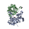
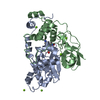
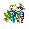
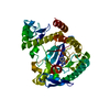
Method to determine structure: MAD / Resolution: 1.9→46.61 Å / Cross valid method: THROUGHOUT / σ(F): 0
Details: THERE IS A SIGNIFICANT PIECE OF DENSITY THAT HAS NOT BEEN ACCOUNTED FOR IN THE MODEL THAT IS LOCATED IN THE PUTATIVE ACTIVE SITE. IT IS "SANDWICHED" BETWEEN HIS 57 AND MSE 60. IT IS ALSO ...Details: THERE IS A SIGNIFICANT PIECE OF DENSITY THAT HAS NOT BEEN ACCOUNTED FOR IN THE MODEL THAT IS LOCATED IN THE PUTATIVE ACTIVE SITE. IT IS "SANDWICHED" BETWEEN HIS 57 AND MSE 60. IT IS ALSO WITHIN INTER-ACTION DISTANCE OF A WATER (HOH 14) THAT IS COORDINATED TO THE ACTIVE SITE METAL ION. IT IS NOT YET POSSIBLE TO ASSIGN THE IDENTITY OF THIS/THESE MOLECULES AS THE STRUCTURE IS OF A PROTEIN OF UNKNOWN FUNCTION. FURTHER BIOCHEMICAL CHARACTERIZATION OF TM0936 MAY AID IN ELUCIDATING THE NATURE OF THIS DENSITY. THE MODEL SEQUENCE MATCHES THE DATABASE SEQUENCE WITH THE FOLLOWING EXCEPTIONS: RESIDUES -1 AND 0 ARE HIS 5 AND 6 OF THE 6-HIS PURIFICATION TAG. THERE IS NO DENSITY FOR SER406 AND THIS DOES NOT APPEAR IN THE MODEL. PROCHECK IDENTIFIES 3 RESIDUES IN DISFAVORED CONFORMATIONS. HIS 228 (DISALLOWED REGION), ASP 279 AND ASN 285 (GENEROUSLY ALLOWED REGION). HIS 228 AND ASP 279 COORDINATE THE PUTATIVE ACTIVE SITE METAL ION. THE BINDING OF THE ACTIVE SITE METAL ION APPEARS TO INDUCE THESE OTHERWISE UNFAVORABLE GEOMETRIES. ASN 285 IS ON THE PROTEIN SURFACE AT A CRYSTALLOGRAPHIC INTERFACE. PROCHECK ADDITIONALLY IDENTIFIES 1 CIS-PEPTIDE RESIDUE PRO 352 WHICH DOES NOT APPEAR TO BE INVOLVED IN THE ACTIVE SITE. A METAL ION IDENTIFIED AS NICKEL BY FLUORESCENCE SCAN IS COORDINATED IN THE PUTATIVE ACTIVE SITE. AS WITH THE UNIDENTIFIED DENSITY PREVIOUSLY NOTED, IT IS NOT POSSIBLE TO DETERMINE IF THE NICKEL ION IS AN ARTIFACT OF NICKEL-CHELATING PURIFICATION OF TM0936 OR THE NATIVE METAL REQUIRED FOR ACTIVITY AS TM0936 IS A PROTEIN OF UNKNOWN FUNCTION. FURTHER BIOCHEMICAL CHARACTERIZATION OF TM0936 MAY AID IN ELUCIDATING THE NATURE OF THIS METAL ION.
RfactorNum. reflection% reflectionSelection details
Rfree0.202 2359 4.9 %RANDOM
Rwork0.175 ---
obs0.175 47228 98.1 %-
Solvent computationSolvent model: BULK SOLVENT CORRECTION / Bsol: 53.8 Å2 / ksol: 0.41 e/Å3
Displacement parametersBiso mean: 24.1 Å2
Baniso -1Baniso -2Baniso -3
1-2.756 Å21.16 Å20 Å2
2--2.774 Å20 Å2
3----5.53 Å2
Refine analyzeLuzzati coordinate error obs: 0.74 Å / Luzzati d res low obs: 6 Å
Refinement stepCycle: LAST / Resolution: 1.9→46.61 Å
ProteinNucleic acidLigandSolventTotal
Num. atoms3235 0 1 174 3410
Refine LS restraints
Refine-IDTypeDev idealDev ideal target
X-RAY DIFFRACTIONc_bond_d0.016
X-RAY DIFFRACTIONc_bond_d_na
X-RAY DIFFRACTIONc_bond_d_prot
X-RAY DIFFRACTIONc_angle_d
X-RAY DIFFRACTIONc_angle_d_na
X-RAY DIFFRACTIONc_angle_d_prot
X-RAY DIFFRACTIONc_angle_deg1.79
X-RAY DIFFRACTIONc_angle_deg_na
X-RAY DIFFRACTIONc_angle_deg_prot
X-RAY DIFFRACTIONc_dihedral_angle_d
X-RAY DIFFRACTIONc_dihedral_angle_d_na
X-RAY DIFFRACTIONc_dihedral_angle_d_prot
X-RAY DIFFRACTIONc_improper_angle_d
X-RAY DIFFRACTIONc_improper_angle_d_na
X-RAY DIFFRACTIONc_improper_angle_d_prot
X-RAY DIFFRACTIONc_mcbond_it3.7491.5
X-RAY DIFFRACTIONc_mcangle_it4.2152
X-RAY DIFFRACTIONc_scbond_it5.7042
X-RAY DIFFRACTIONc_scangle_it6.9532.5
LS refinement shellResolution: 1.9→1.99 Å
RfactorNum. reflection% reflection
Rfree0.2534 265 5.1 %
Rwork0.2327 4845 -

+
About Yorodumi

-
News

-
Feb 9, 2022. New format data for meta-information of EMDB entries

New format data for meta-information of EMDB entries

  • Version 3 of the EMDB header file is now the official format.
  • The previous official version 1.9 will be removed from the archive.

Related info.:EMDB header

External links:wwPDB to switch to version 3 of the EMDB data model

-
Aug 12, 2020. Covid-19 info

Covid-19 info

URL: https://pdbjlc1.pdbj.org/emnavi/covid19.php

New page: Covid-19 featured information page in EM Navigator.

Related info.:Covid-19 info / Mar 5, 2020. Novel coronavirus structure data

+
Mar 5, 2020. Novel coronavirus structure data

Novel coronavirus structure data

Related info.:Yorodumi Speices / Aug 12, 2020. Covid-19 info

External links:COVID-19 featured content - PDBj / Molecule of the Month (242):Coronavirus Proteases

+
Jan 31, 2019. EMDB accession codes are about to change! (news from PDBe EMDB page)

EMDB accession codes are about to change! (news from PDBe EMDB page)

  • The allocation of 4 digits for EMDB accession codes will soon come to an end. Whilst these codes will remain in use, new EMDB accession codes will include an additional digit and will expand incrementally as the available range of codes is exhausted. The current 4-digit format prefixed with “EMD-” (i.e. EMD-XXXX) will advance to a 5-digit format (i.e. EMD-XXXXX), and so on. It is currently estimated that the 4-digit codes will be depleted around Spring 2019, at which point the 5-digit format will come into force.
  • The EM Navigator/Yorodumi systems omit the EMD- prefix.

Related info.:Q: What is EMD? / ID/Accession-code notation in Yorodumi/EM Navigator

External links:EMDB Accession Codes are Changing Soon! / Contact to PDBj

+
Jul 12, 2017. Major update of PDB

Major update of PDB

  • wwPDB released updated PDB data conforming to the new PDBx/mmCIF dictionary.
  • This is a major update changing the version number from 4 to 5, and with Remediation, in which all the entries are updated.
  • In this update, many items about electron microscopy experimental information are reorganized (e.g. em_software).
  • Now, EM Navigator and Yorodumi are based on the updated data.

External links:wwPDB Remediation / Enriched Model Files Conforming to OneDep Data Standards Now Available in the PDB FTP Archive

-
Yorodumi

Thousand views of thousand structures

  • Yorodumi is a browser for structure data from EMDB, PDB, SASBDB, etc.
  • This page is also the successor to EM Navigator detail page, and also detail information page/front-end page for Omokage search.
  • The word "yorodu" (or yorozu) is an old Japanese word meaning "ten thousand". "mi" (miru) is to see.

Related info.:EMDB / PDB / SASBDB / Comparison of 3 databanks / Yorodumi Search / Aug 31, 2016. New EM Navigator & Yorodumi / Yorodumi Papers / Jmol/JSmol / Function and homology information / Changes in new EM Navigator and Yorodumi

Read more