 Movie
Movie Controller

[English] 日本語
 Yorodumi
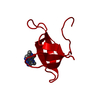
- PDB-2p4t: Structure of the Q67H mutant of R67 dihydrofolate reductase-NADP+... -
+ Open data

- Basic information
| Entry | Database: PDB / ID: 2p4t | ||||||
|---|---|---|---|---|---|---|---|
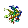
| Title | Structure of the Q67H mutant of R67 dihydrofolate reductase-NADP+ complex reveals a novel cofactor binding mode | ||||||
 Components Components | Dihydrofolate reductase type 2 | ||||||
 Keywords Keywords | OXIDOREDUCTASE / bacterial infections / folate metabolism / NADP+ / R67 DHFR / Symmetric binding / trimethoprim-resistance | ||||||
| Function / homology |  Function and homology information Function and homology informationresponse to methotrexate / dihydrofolate reductase / dihydrofolate reductase activity / tetrahydrofolate biosynthetic process / one-carbon metabolic process / response to xenobiotic stimulus / response to antibiotic Similarity search - Function | ||||||
| Biological species |  | ||||||
| Method |  X-RAY DIFFRACTION / SYNCHROTRON / MOLECULAR REPLACEMENT / Resolution: 1.15 Å X-RAY DIFFRACTION / SYNCHROTRON / MOLECULAR REPLACEMENT / Resolution: 1.15 Å | ||||||
 Authors Authors | Divya, N. / Grifith, E. / Narayana, N. | ||||||
 Citation Citation | Journal: Protein Sci. / Year: 2007 Title: Structure of the Q67H mutant of R67 dihydrofolate reductase-NADP+ complex reveals a novel cofactor binding mode. Authors: Divya, N. / Grifith, E. / Narayana, N. #1: Journal: Acta Crystallogr.,Sect.D / Year: 2006Title: High-resolution structure of a plasmid-encoded dihydrofolate reductase: pentagonal network of water molecules in the D2-symmetric active site Authors: Narayana, N. #2: Journal: Nat.Struct.Biol. / Year: 1995Title: A Plasmid-Encoded Dihydrofolate Reductase from Trimethoprim-Resistant Bacteria has a Novel D2-Symmetric Active Site Authors: Narayana, N. / Matthews, D.A. / Howell, E.E. / Xuong, N. #3: Journal: Protein Eng. / Year: 1997 Title: A glutamine 67-->histidine mutation in homotetrameric R67 dihydrofolate reductase results in four mutations per single active site pore and causes substantial substrate and cofactor inhibition Authors: Park, H. / Bradrick, T.D. / Howell, E.E. | ||||||
| History |
|
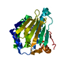
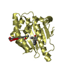
- Structure visualization
| Structure viewer | Molecule: MolmilJmol/JSmol |
|---|
- Downloads & links
-Download
| PDBx/mmCIF format | 2p4t.cif.gz | 28.5 KB | Display | PDBx/mmCIF format |
|---|---|---|---|---|
| PDB format | pdb2p4t.ent.gz | 17.1 KB | Display | PDB format |
| PDBx/mmJSON format | 2p4t.json.gz | Tree view | PDBx/mmJSON format | |
| Others |  Other downloads Other downloads |
-Validation report
| Arichive directory | https://data.pdbj.org/pub/pdb/validation_reports/p4/2p4tftp://data.pdbj.org/pub/pdb/validation_reports/p4/2p4t | HTTPS FTP |
|---|
-Related structure data
| Related structure data |  1vieS S: Starting model for refinement |
|---|---|
| Similar structure data |


-Links
 PDBj
PDBj


- Assembly
| Deposited unit | 
| ||||||||
|---|---|---|---|---|---|---|---|---|---|
| 1 | 
| ||||||||
| Unit cell |
| ||||||||
| Details | The biological assembly is comprised of four subunits generated by symmetry axes. The crystallographic 222 symmetry generates the biologically active tetramer. x,y,z; -x+1,-y+1,z; y,x,-z+1; -y+1,-x+1,-z+1 |
-Components
| #1: Protein | Mass: 6742.546 Da / Num. of mol.: 1 / Mutation: Residues 1-16 are cleaved and Q67H mutation Source method: isolated from a genetically manipulated source Source: (gene. exp.) Strain: TMP-RESISTANT, CONTAINING R67 DHFR OVERPRODUCING PLASMID PLZ1 Production host: |
|---|---|
| #2: Chemical | ChemComp-NAP /   Mass: 743.405 Da / Num. of mol.: 1 / Source method: obtained synthetically / Formula: C21H28N7O17P3 Mass: 743.405 Da / Num. of mol.: 1 / Source method: obtained synthetically / Formula: C21H28N7O17P3 |
| #3: Water | ChemComp-HOH /  Mass: 18.015 Da / Num. of mol.: 78 / Source method: isolated from a natural source / Formula: H2O Mass: 18.015 Da / Num. of mol.: 78 / Source method: isolated from a natural source / Formula: H2O |
-Experimental details
-Experiment
| Experiment | Method: X-RAY DIFFRACTION / Number of used crystals: 1 |
|---|
- Sample preparation
| Crystal | Density Matthews: 2.23 Å3/Da / Density % sol: 44.81 % |
|---|---|
| Crystal grow | Temperature: 277 K / Method: vapor diffusion, hanging drop / pH: 7.5 Details: 0.1M Tris-Hcl buffer, 20% PEG1000 and 10% MPD, pH 7.5, VAPOR DIFFUSION, HANGING DROP, temperature 277K |
-Data collection
| Diffraction | Mean temperature: 100 K |
|---|---|
| Diffraction source | Source: SYNCHROTRON / Site: CHESS  / Beamline: F1 / Wavelength: 0.9792 Å / Beamline: F1 / Wavelength: 0.9792 Å |
| Detector | Type: ADSC QUANTUM 210 / Detector: CCD / Date: Apr 23, 2005 |
| Radiation | Protocol: SINGLE WAVELENGTH / Monochromatic (M) / Laue (L): M / Scattering type: x-ray |
| Radiation wavelength | Wavelength: 0.9792 Å / Relative weight: 1 |
| Reflection | Resolution: 1.15→20 Å / Num. all: 19048 / Num. obs: 18488 / % possible obs: 86.6 % / Observed criterion σ(I): 1 / Redundancy: 7.1 % / Rsym value: 0.039 / Net I/σ(I): 30.2 |
| Reflection shell | Resolution: 1.15→1.24 Å / Redundancy: 1.4 % / Mean I/σ(I) obs: 2.4 / Num. unique all: 1609 / Rsym value: 0.285 / % possible all: 60.2 |
- Processing
| Software |
| |||||||||||||||||||||||||
|---|---|---|---|---|---|---|---|---|---|---|---|---|---|---|---|---|---|---|---|---|---|---|---|---|---|---|
| Refinement | Method to determine structure: MOLECULAR REPLACEMENT Starting model: PDB entry 1VIE Resolution: 1.15→20 Å / Cross valid method: THROUGHOUT / σ(F): 1 / σ(I): 1 / Stereochemistry target values: Engh & Huber Details: HOH 149 HAS PARTIAL OCCUPANCY. WHEN COFACTOR IS BOUND, THE HOH 149 IS ABSENT. HOH 146 ALSO HAS PARTIAL OCCUPANCY.THE ADENOSINE PHOSPHATE PORTION OF THE COFACTOR IS NOT SEEN IN THE DENSITY. ...Details: HOH 149 HAS PARTIAL OCCUPANCY. WHEN COFACTOR IS BOUND, THE HOH 149 IS ABSENT. HOH 146 ALSO HAS PARTIAL OCCUPANCY.THE ADENOSINE PHOSPHATE PORTION OF THE COFACTOR IS NOT SEEN IN THE DENSITY. THEREFORE, THE RESPECTIVE ATOMIC COORDINATES ARE MISSING IN THIS LIST.
| |||||||||||||||||||||||||
| Refinement step | Cycle: LAST / Resolution: 1.15→20 Å
| |||||||||||||||||||||||||
| LS refinement shell | Resolution: 1.15→1.24 Å
|
