 Movie
Movie Controller

[English] 日本語
 Yorodumi
- PDB-2gqv: High-resolution structure of a plasmid-encoded dihydrofolate redu... -
+ Open data

- Basic information
| Entry | Database: PDB / ID: 2gqv | ||||||
|---|---|---|---|---|---|---|---|
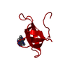
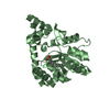
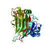
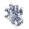
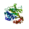
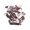
| Title | High-resolution structure of a plasmid-encoded dihydrofolate reductase: pentagonal network of water molecules in the D2-symmetric active site | ||||||
 Components Components | Dihydrofolate reductase type 2 | ||||||
 Keywords Keywords | OXIDOREDUCTASE / anisotropic refinement / atomic-resolution structure / folate metabolism / plasmid-encoded R67 DHFR / TMP-resistant DHFR | ||||||
| Function / homology |  Function and homology information Function and homology informationresponse to methotrexate / dihydrofolate reductase / dihydrofolate reductase activity / tetrahydrofolate biosynthetic process / one-carbon metabolic process / response to xenobiotic stimulus / response to antibiotic Similarity search - Function | ||||||
| Biological species |  | ||||||
| Method |  X-RAY DIFFRACTION / SYNCHROTRON / MOLECULAR REPLACEMENT / Resolution: 1.1 Å X-RAY DIFFRACTION / SYNCHROTRON / MOLECULAR REPLACEMENT / Resolution: 1.1 Å | ||||||
 Authors Authors | Narayana, N. | ||||||
 Citation Citation | Journal: Acta Crystallogr.,Sect.D / Year: 2006 Title: High-resolution structure of a plasmid-encoded dihydrofolate reductase: pentagonal network of water molecules in the D(2)-symmetric active site. Authors: Narayana, N. #1: Journal: Nat.Struct.Biol. / Year: 1995Title: A plasmid-encoded dihydrofolate reductase from trimethoprim-resistant bacteria has a novel D2-symmetric active site. Authors: Narayana, N. / Matthews, D.A. / Howell, E.E. / Nguyen-huu, X. #2: Journal: Biochemistry / Year: 1986 Title: Crystal structure of a novel trimethoprim-resistant dihydrofolate reductase specified in Escherichia coli by R-plasmid R67. Authors: Matthews, D.A. / Smith, S.L. / Baccanari, D.P. / Burchall, J.J. / Oatley, S.J. / Kraut, J. | ||||||
| History |
|
- Structure visualization
| Structure viewer | Molecule: MolmilJmol/JSmol |
|---|
- Downloads & links
-Download
| PDBx/mmCIF format | 2gqv.cif.gz | 53.6 KB | Display | PDBx/mmCIF format |
|---|---|---|---|---|
| PDB format | pdb2gqv.ent.gz | 39 KB | Display | PDB format |
| PDBx/mmJSON format | 2gqv.json.gz | Tree view | PDBx/mmJSON format | |
| Others |  Other downloads Other downloads |
-Validation report
| Arichive directory | https://data.pdbj.org/pub/pdb/validation_reports/gq/2gqvftp://data.pdbj.org/pub/pdb/validation_reports/gq/2gqv | HTTPS FTP |
|---|
-Related structure data
| Related structure data |  1vieS S: Starting model for refinement |
|---|---|
| Similar structure data |


-Links
 PDBj
PDBj


- Assembly
| Deposited unit | 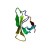
| ||||||||||||
|---|---|---|---|---|---|---|---|---|---|---|---|---|---|
| 1 | 
| ||||||||||||
| Unit cell |
| ||||||||||||
| Components on special symmetry positions |
| ||||||||||||
| Details | The biological assembly is comprised of four subunits generated by symmetry axes. The crystallographic 222 symmetry generates the biologically active tetramer. x,y,z; -x+1,-y+1,z; y,x,-z+1; -y+1,-x+1,-z+1 |

-Components
| #1: Protein | Mass: 6732.528 Da / Num. of mol.: 1 Source method: isolated from a genetically manipulated source Source: (gene. exp.) Strain: TMP-RESISTANT, CONTAINING R67 DHFR OVERPRODUCING PLASMID PLZ1 Production host: |
|---|---|
| #2: Water | ChemComp-HOH /  Mass: 18.015 Da / Num. of mol.: 191 / Source method: isolated from a natural source / Formula: H2O Mass: 18.015 Da / Num. of mol.: 191 / Source method: isolated from a natural source / Formula: H2O |
-Experimental details
-Experiment
| Experiment | Method: X-RAY DIFFRACTION / Number of used crystals: 1 |
|---|
- Sample preparation
| Crystal | Density Matthews: 2.24 Å3/Da / Density % sol: 45.2 % |
|---|---|
| Crystal grow | Temperature: 277 K / Method: vapor diffusion, hanging drop / pH: 7.5 Details: 0.1M Tris-Hcl buffer, 25% MPD, pH 7.5, VAPOR DIFFUSION, HANGING DROP, temperature 277K |
-Data collection
| Diffraction | Mean temperature: 100 K |
|---|---|
| Diffraction source | Source: SYNCHROTRON / Site: APS  / Beamline: 14-BM-D / Wavelength: 0.9495 Å / Beamline: 14-BM-D / Wavelength: 0.9495 Å |
| Detector | Type: ADSC QUANTUM 1 / Detector: CCD / Date: Mar 15, 1998 |
| Radiation | Protocol: SINGLE WAVELENGTH / Monochromatic (M) / Laue (L): M / Scattering type: x-ray |
| Radiation wavelength | Wavelength: 0.9495 Å / Relative weight: 1 |
| Reflection | Resolution: 1.1→50 Å / Num. all: 22453 / Num. obs: 19914 / % possible obs: 90 % / Observed criterion σ(F): 2 / Observed criterion σ(I): 4 / Redundancy: 8.5 % / Biso Wilson estimate: 9.7 Å2 / Rsym value: 0.042 / Net I/σ(I): 21.4 |
| Reflection shell | Resolution: 1.1→1.14 Å / Rsym value: 0.34 / % possible all: 58 |
- Processing
| Software |
| |||||||||||||||||||||||||||||||||
|---|---|---|---|---|---|---|---|---|---|---|---|---|---|---|---|---|---|---|---|---|---|---|---|---|---|---|---|---|---|---|---|---|---|---|
| Refinement | Method to determine structure: MOLECULAR REPLACEMENT Starting model: PDB ENTRY 1VIE Resolution: 1.1→10 Å / Cross valid method: THROUGHOUT / σ(F): 0 / σ(I): 0 / Stereochemistry target values: Engh & Huber
| |||||||||||||||||||||||||||||||||
| Solvent computation | Solvent model: MOEWS & KRETSINGER, J.MOL.BIOL.91(1973)201-228 | |||||||||||||||||||||||||||||||||
| Refinement step | Cycle: LAST / Resolution: 1.1→10 Å
| |||||||||||||||||||||||||||||||||
| Refine LS restraints |
| |||||||||||||||||||||||||||||||||
| LS refinement shell | Highest resolution: 1.1 Å / Rfactor Rfree error: 0.012
|
