 Movie
Movie Controller

[English] 日本語
 Yorodumi
- PDB-2vfb: The structure of Mycobacterium marinum arylamine N-acetyltransferase -
+ Open data

- Basic information
| Entry | Database: PDB / ID: 2vfb | ||||||
|---|---|---|---|---|---|---|---|
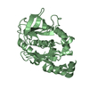
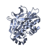
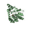
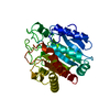
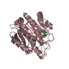
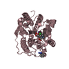
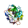
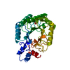
| Title | The structure of Mycobacterium marinum arylamine N-acetyltransferase | ||||||
 Components Components | ARYLAMINE N-ACETYLTRANSFERASE | ||||||
 Keywords Keywords | TRANSFERASE / ARYLAMINE N-ACETYLTRANSFERASE / NAT / ACETYL COA / MYCOBACTERIA / ACTYLTRANSFERASE | ||||||
| Function / homology |  Function and homology information Function and homology information | ||||||
| Biological species |  MYCOBACTERIUM MARINUM (bacteria) MYCOBACTERIUM MARINUM (bacteria) | ||||||
| Method |  X-RAY DIFFRACTION / SYNCHROTRON / MOLECULAR REPLACEMENT / Resolution: 2 Å X-RAY DIFFRACTION / SYNCHROTRON / MOLECULAR REPLACEMENT / Resolution: 2 Å | ||||||
 Authors Authors | Fullam, E. / Westwood, I.M. / Anderton, M.C. / Lowe, E.D. / Sim, E. / Noble, M.E.M. | ||||||
 Citation Citation | Journal: J.Mol.Biol. / Year: 2008 Title: Divergence of Cofactor Recognition Across Evolution: Coenzyme a Binding in a Prokaryotic Arylamine N-Acetyltransferase. Authors: Fullam, E. / Westwood, I.M. / Anderton, M.C. / Lowe, E.D. / Sim, E. / Noble, M.E.M. | ||||||
| History |
|
- Structure visualization
| Structure viewer | Molecule: MolmilJmol/JSmol |
|---|
- Downloads & links
-Download
| PDBx/mmCIF format | 2vfb.cif.gz | 67.9 KB | Display | PDBx/mmCIF format |
|---|---|---|---|---|
| PDB format | pdb2vfb.ent.gz | 49.7 KB | Display | PDB format |
| PDBx/mmJSON format | 2vfb.json.gz | Tree view | PDBx/mmJSON format | |
| Others |  Other downloads Other downloads |
-Validation report
| Arichive directory | https://data.pdbj.org/pub/pdb/validation_reports/vf/2vfbftp://data.pdbj.org/pub/pdb/validation_reports/vf/2vfb | HTTPS FTP |
|---|
-Related structure data
| Related structure data | 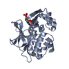 2vfcC 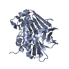 1gx3S C: citing same article ( S: Starting model for refinement |
|---|---|
| Similar structure data |


-Links
 PDBj
PDBj- Assembly
| Deposited unit | 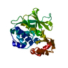
| ||||||||
|---|---|---|---|---|---|---|---|---|---|
| 1 |
| ||||||||
| Unit cell |
|
-Components
| #1: Protein | Mass: 30668.752 Da / Num. of mol.: 1 Source method: isolated from a genetically manipulated source Source: (gene. exp.) MYCOBACTERIUM MARINUM (bacteria) / Production host: References: UniProt: B2HIZ6*PLUS, arylamine N-acetyltransferase |
|---|---|
| #2: Water | ChemComp-HOH /  Mass: 18.015 Da / Num. of mol.: 139 / Source method: isolated from a natural source / Formula: H2O Mass: 18.015 Da / Num. of mol.: 139 / Source method: isolated from a natural source / Formula: H2O |
| Sequence details | C-TERMINAL HIS TAG ADDED |
-Experimental details
-Experiment
| Experiment | Method: X-RAY DIFFRACTION / Number of used crystals: 1 |
|---|
- Sample preparation
| Crystal | Density Matthews: 1.86 Å3/Da / Density % sol: 33.9 % / Description: NONE |
|---|---|
| Crystal grow | pH: 7.5 Details: 200MM CACL2, 100MM HEPES, PH7.5, 28% POLY(ETHYLENE GLYCOL) 400 |
-Data collection
| Diffraction | Mean temperature: 100 K |
|---|---|
| Diffraction source | Source: SYNCHROTRON / Site: ESRF  / Beamline: ID29 / Wavelength: 0.98 / Beamline: ID29 / Wavelength: 0.98 |
| Detector | Type: ADSC CCD / Detector: CCD / Details: TOROIDAL MIRROR |
| Radiation | Monochromator: SI(111) / Protocol: SINGLE WAVELENGTH / Monochromatic (M) / Laue (L): M / Scattering type: x-ray |
| Radiation wavelength | Wavelength: 0.98 Å / Relative weight: 1 |
| Reflection | Resolution: 1.45→51.5 Å / Num. obs: 16884 / % possible obs: 100 % / Observed criterion σ(I): 0 / Redundancy: 8.9 % / Rmerge(I) obs: 0.09 / Net I/σ(I): 6.7 |
| Reflection shell | Resolution: 2→2.11 Å / Redundancy: 9.24 % / Rmerge(I) obs: 0.48 / Mean I/σ(I) obs: 1.6 / % possible all: 100 |
- Processing
| Software |
| ||||||||||||||||||||||||||||||||||||||||||||||||||||||||||||||||||||||||||||||||||||||||||||||||||||||||||||||||||||||||||||||||||||||||||||||||||||||||||||||||||||||||||||||||||||||
|---|---|---|---|---|---|---|---|---|---|---|---|---|---|---|---|---|---|---|---|---|---|---|---|---|---|---|---|---|---|---|---|---|---|---|---|---|---|---|---|---|---|---|---|---|---|---|---|---|---|---|---|---|---|---|---|---|---|---|---|---|---|---|---|---|---|---|---|---|---|---|---|---|---|---|---|---|---|---|---|---|---|---|---|---|---|---|---|---|---|---|---|---|---|---|---|---|---|---|---|---|---|---|---|---|---|---|---|---|---|---|---|---|---|---|---|---|---|---|---|---|---|---|---|---|---|---|---|---|---|---|---|---|---|---|---|---|---|---|---|---|---|---|---|---|---|---|---|---|---|---|---|---|---|---|---|---|---|---|---|---|---|---|---|---|---|---|---|---|---|---|---|---|---|---|---|---|---|---|---|---|---|---|---|
| Refinement | Method to determine structure: MOLECULAR REPLACEMENT Starting model: PDB ENTRY 1GX3 Resolution: 2→29.06 Å / Cor.coef. Fo:Fc: 0.948 / Cor.coef. Fo:Fc free: 0.931 / SU B: 9.934 / SU ML: 0.127 / TLS residual ADP flag: LIKELY RESIDUAL / Cross valid method: THROUGHOUT / ESU R: 0.215 / ESU R Free: 0.178 / Stereochemistry target values: MAXIMUM LIKELIHOOD / Details: HYDROGENS HAVE BEEN ADDED IN THE RIDING POSITIONS.
| ||||||||||||||||||||||||||||||||||||||||||||||||||||||||||||||||||||||||||||||||||||||||||||||||||||||||||||||||||||||||||||||||||||||||||||||||||||||||||||||||||||||||||||||||||||||
| Solvent computation | Ion probe radii: 0.8 Å / Shrinkage radii: 0.8 Å / VDW probe radii: 1.4 Å / Solvent model: MASK | ||||||||||||||||||||||||||||||||||||||||||||||||||||||||||||||||||||||||||||||||||||||||||||||||||||||||||||||||||||||||||||||||||||||||||||||||||||||||||||||||||||||||||||||||||||||
| Displacement parameters | Biso mean: 23.98 Å2
| ||||||||||||||||||||||||||||||||||||||||||||||||||||||||||||||||||||||||||||||||||||||||||||||||||||||||||||||||||||||||||||||||||||||||||||||||||||||||||||||||||||||||||||||||||||||
| Refinement step | Cycle: LAST / Resolution: 2→29.06 Å
| ||||||||||||||||||||||||||||||||||||||||||||||||||||||||||||||||||||||||||||||||||||||||||||||||||||||||||||||||||||||||||||||||||||||||||||||||||||||||||||||||||||||||||||||||||||||
| Refine LS restraints |
|