 Movie
Movie Controller

[English] 日本語
 Yorodumi
- PDB-1tml: CRYSTAL STRUCTURE OF THE CATALYTIC DOMAIN OF A THERMOPHILIC ENDOC... -
+ Open data

- Basic information
| Entry | Database: PDB / ID: 1tml | ||||||
|---|---|---|---|---|---|---|---|
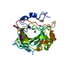
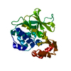
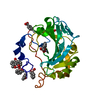
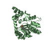
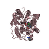
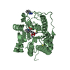
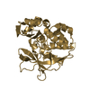
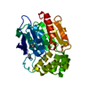
| Title | CRYSTAL STRUCTURE OF THE CATALYTIC DOMAIN OF A THERMOPHILIC ENDOCELLULASE | ||||||
 Components Components | ENDO-1,4-BETA-D-GLUCANASE | ||||||
 Keywords Keywords | BETA-AMYLASE / ENDOCELLULASE / CATALYTIC DOMAIN | ||||||
| Function / homology |  Function and homology information Function and homology informationcellulase / cellulase activity / polysaccharide binding / cellulose catabolic process Similarity search - Function | ||||||
| Biological species |   Thermobifida fusca (bacteria) Thermobifida fusca (bacteria) | ||||||
| Method |  X-RAY DIFFRACTION / Resolution: 1.8 Å X-RAY DIFFRACTION / Resolution: 1.8 Å | ||||||
 Authors Authors | Spezio, M. / Wilson, D.B. / Karplus, P.A. | ||||||
 Citation Citation | Journal: Biochemistry / Year: 1993 Title: Crystal structure of the catalytic domain of a thermophilic endocellulase. Authors: Spezio, M. / Wilson, D.B. / Karplus, P.A. #1: Journal: To be PublishedTitle: Crystal Structure of the Catalytic Domain of a Thermophilic Endocellulase Authors: Spezio, M. / Wilson, D.B. / Karplus, P.A. | ||||||
| History |
| ||||||
| Remark 700 | SHEET THE SHEET PRESENTED AS *COR* ON SHEET RECORDS BELOW IS ACTUALLY AN EIGHT STRANDED BETA-BARREL. ...SHEET THE SHEET PRESENTED AS *COR* ON SHEET RECORDS BELOW IS ACTUALLY AN EIGHT STRANDED BETA-BARREL. THIS IS REPRESENTED BY A NINE-STRANDED SHEET IN WHICH THE FIRST AND LAST STRANDS ARE IDENTICAL. IN ADDITION, STRAND 1 IS BIFURCATED THUS THE BARREL IS REPRESENTED BY TWO SETS OF SHEET RECORDS *COR* AND *BOR* WHERE STRANDS 2, 3, 4, 5, 6, 7, AND 8 ARE IDENTICAL. |
- Structure visualization
| Structure viewer | Molecule: MolmilJmol/JSmol |
|---|
- Downloads & links
-Download
| PDBx/mmCIF format | 1tml.cif.gz | 79.3 KB | Display | PDBx/mmCIF format |
|---|---|---|---|---|
| PDB format | pdb1tml.ent.gz | 60 KB | Display | PDB format |
| PDBx/mmJSON format | 1tml.json.gz | Tree view | PDBx/mmJSON format | |
| Others |  Other downloads Other downloads |
-Validation report
| Arichive directory | https://data.pdbj.org/pub/pdb/validation_reports/tm/1tmlftp://data.pdbj.org/pub/pdb/validation_reports/tm/1tml | HTTPS FTP |
|---|
-Related structure data
| Similar structure data |
|---|


-Links
 PDBj
PDBj


- Assembly
| Deposited unit | 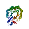
| ||||||||
|---|---|---|---|---|---|---|---|---|---|
| 1 |
| ||||||||
| Unit cell |
| ||||||||
| Atom site foot note | 1: RESIDUES ASN 1 AND ASP 2 ARE NOT WELL ORDERED AND HAVE VERY WEAK ELECTRON DENSITY. 2: RESIDUE TRP 162, IN THE ACTIVE SITE CLEFT, IS WELL ORDERED AND HAS STRONG DENSITY EVEN THOUGH ITS MAIN CHAIN TORSION ANGLES ARE OUTSIDE THE FAVORABLE REGIONS OF A RAMACHANDRAN PLOT. |
-Components
| #1: Protein | Mass: 30436.793 Da / Num. of mol.: 1 Source method: isolated from a genetically manipulated source Source: (gene. exp.) Thermobifida fusca (bacteria) / References: UniProt: P26222, cellulase |
|---|---|
| #2: Chemical | ChemComp-SO4 /   Mass: 96.063 Da / Num. of mol.: 1 / Source method: obtained synthetically / Formula: SO4 Mass: 96.063 Da / Num. of mol.: 1 / Source method: obtained synthetically / Formula: SO4 |
| #3: Water | ChemComp-HOH /  Mass: 18.015 Da / Num. of mol.: 73 / Source method: isolated from a natural source / Formula: H2O Mass: 18.015 Da / Num. of mol.: 73 / Source method: isolated from a natural source / Formula: H2O |
| Compound details | D1 AND F1 OF THE HELIX ARE SMALL AND ARE NOT EXACTLY CONSERVED BETWEEN THIS STRUCTURE AND ...D1 AND F1 OF THE HELIX ARE SMALL AND ARE NOT EXACTLY CONSERVED BETWEEN THIS STRUCTURE AND CELLOBIOHY |
| Has protein modification | Y |
| Sequence details | SEQUENCE ADVISORY NOTICE: DIFFERENCE BETWEEN SWISS-PROT AND PDB SEQUENCE. SWISS-PROT ENTRY NAME: ...SEQUENCE ADVISORY NOTICE: DIFFERENCE |
-Experimental details
-Experiment
| Experiment | Method: X-RAY DIFFRACTION |
|---|
- Sample preparation
| Crystal | Density Matthews: 1.94 Å3/Da / Density % sol: 36.62 % | ||||||||||||||||||||||||||||||||||||||||||
|---|---|---|---|---|---|---|---|---|---|---|---|---|---|---|---|---|---|---|---|---|---|---|---|---|---|---|---|---|---|---|---|---|---|---|---|---|---|---|---|---|---|---|---|
| Crystal grow | *PLUS pH: 6 / Method: vapor diffusion, hanging drop | ||||||||||||||||||||||||||||||||||||||||||
| Components of the solutions | *PLUS
|
-Data collection
| Reflection | *PLUS Highest resolution: 1.8 Å / Num. obs: 21081 / % possible obs: 97 % / Num. measured all: 79576 / Rmerge(I) obs: 0.0459 |
|---|
- Processing
| Software |
| ||||||||||||||||||||||||||||||||||||||||||||||||||||||||||||
|---|---|---|---|---|---|---|---|---|---|---|---|---|---|---|---|---|---|---|---|---|---|---|---|---|---|---|---|---|---|---|---|---|---|---|---|---|---|---|---|---|---|---|---|---|---|---|---|---|---|---|---|---|---|---|---|---|---|---|---|---|---|
| Refinement | Rfactor Rwork: 0.184 / Rfactor obs: 0.184 / Highest resolution: 1.8 Å Details: RESIDUES ASN 1 AND ASP 2 ARE NOT WELL ORDERED AND HAVE VERY WEAK ELECTRON DENSITY. RESIDUE TRP 162, IN THE ACTIVE SITE CLEFT, IS WELL ORDERED AND HAS STRONG DENSITY EVEN THOUGH ITS MAIN ...Details: RESIDUES ASN 1 AND ASP 2 ARE NOT WELL ORDERED AND HAVE VERY WEAK ELECTRON DENSITY. RESIDUE TRP 162, IN THE ACTIVE SITE CLEFT, IS WELL ORDERED AND HAS STRONG DENSITY EVEN THOUGH ITS MAIN CHAIN TORSION ANGLES ARE OUTSIDE THE FAVORABLE REGIONS OF A RAMACHANDRAN PLOT. | ||||||||||||||||||||||||||||||||||||||||||||||||||||||||||||
| Refinement step | Cycle: LAST / Highest resolution: 1.8 Å
| ||||||||||||||||||||||||||||||||||||||||||||||||||||||||||||
| Refine LS restraints |
|
