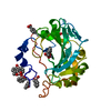
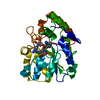
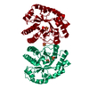
- PDB-1vpq: CRYSTAL STRUCTURE OF a DUF72 family protein (TM1631) FROM THERMOT... -
+
Open data
ID or keywords:
Loading...
-
Basic information
Entry
Database: PDB / ID: 1vpq
Title
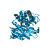
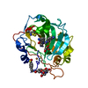
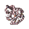
CRYSTAL STRUCTURE OF a DUF72 family protein (TM1631) FROM THERMOTOGA MARITIMA MSB8 AT 2.20 A RESOLUTION
Components
hypothetical protein TM1631
Keywords
UNKNOWN FUNCTION / STRUCTURAL GENOMICS / JOINT CENTER FOR STRUCTURAL GENOMICS / JCSG / PROTEIN STRUCTURE INITIATIVE / PSI
Function / homology
Protein of unknown function UPF0759 / Protein of unknown function DUF72 / UPF0759 superfamily / Protein of unknown function DUF72 / TIM Barrel / Alpha-Beta Barrel / Alpha Beta / DUF72 domain-containing protein
Mass: 18.015 Da / Num. of mol.: 91 / Source method: isolated from a natural source / Formula: H2O
-
Experimental details
-
Experiment
Experiment
Method: X-RAY DIFFRACTION / Number of used crystals: 2
-
Sample preparation
Crystal
ID
Density Matthews (Å3/Da)
Density % sol (%)
Description
1
2.2
44.02
2
2.28
45.59
DATA FROM A SE-MET CONTAINING CRYSTAL IN SPACEGROUP P32 WAS USED FOR THE MAD PHASING EXPERIMENTS AT 2.8 ANGSTROMS RESOLUTION. THIS MAD STRUCTURE WAS USED AS A MOLECULAR REPLACEMENT MODEL TO PHASE THE STRUCTURE AT A RESOLUTION OF 2.2 ANGSTROMS IN THE C222(1) SPACEGROUP.
0.2M K3Citrate, 20.0% PEG-3350, No Buffer pH 8.3, VAPOR DIFFUSION,SITTING DROP,NANODROP, temperature 277K
-
Data collection
Diffraction
ID
Mean temperature (K)
Crystal-ID
1
100
1
2
100
1
1,2
1
Diffraction source
Source
Site
Beamline
ID
Wavelength
Wavelength (Å)
SYNCHROTRON
SSRL
BL11-1
1
1.033169
SYNCHROTRON
APS
19-BM
2
0.98036, 0.98014, 0.96427
Detector
Type
ID
Detector
Date
Details (eV)
ADSC QUANTUM 315
1
CCD
Feb 7, 2002
flatmirror
APS
2
CCD
Nov 15, 2003
Rosenbaum-Rock vertical focusing mirror
Radiation
ID
Monochromator
Protocol
Monochromatic (M) / Laue (L)
Scattering type
Wavelength-ID
1
singlecrystalSi(311) bentmonochromator
SINGLEWAVELENGTH
M
x-ray
1
2
Rosenbaum-Rock double-crystal monochromator
MAD
M
x-ray
1
Radiation wavelength
ID
Wavelength (Å)
Relative weight
1
1.033169
1
2
0.98036
1
3
0.98014
1
4
0.96427
1
Reflection
Resolution: 2.2→49.63 Å / Num. obs: 13378 / % possible obs: 88.4 % / Redundancy: 3.2 % / Biso Wilson estimate: 37.6 Å2 / Rmerge(I) obs: 0.095 / Net I/σ(I): 10.5
Reflection shell
Resolution: 2.2→2.26 Å / Redundancy: 2.2 % / Rmerge(I) obs: 0.4 / Mean I/σ(I) obs: 2.8 / Num. unique all: 736 / % possible all: 67.3
-
Processing
Software
Name
Version
Classification
MOSFLM
datareduction
SCALA
4.2)
datascaling
SHARP
phasing
REFMAC
5.2.0005
refinement
CCP4
(SCALA)
datascaling
Refinement
Method to determine structure: MAD, MOLECULAR REPLACEMENT / Resolution: 2.2→49.63 Å / Cor.coef. Fo:Fc: 0.944 / Cor.coef. Fo:Fc free: 0.918 / SU B: 18.974 / SU ML: 0.228 / TLS residual ADP flag: LIKELY RESIDUAL / Cross valid method: THROUGHOUT / ESU R: 0.386 / ESU R Free: 0.26 / Stereochemistry target values: MAXIMUM LIKELIHOOD Details: 1).HYDROGENS HAVE BEEN ADDED IN THE RIDING POSITIONS 2).A SO4 ANION IS LOCATED AT THE TWO-FOLD CRYSTALLOGRAPHIC SYMMETRY AXIS AT THE INTERFACE BETWEEN TWO MONOMERS. THE S AND O ATOMS ON ...Details: 1).HYDROGENS HAVE BEEN ADDED IN THE RIDING POSITIONS 2).A SO4 ANION IS LOCATED AT THE TWO-FOLD CRYSTALLOGRAPHIC SYMMETRY AXIS AT THE INTERFACE BETWEEN TWO MONOMERS. THE S AND O ATOMS ON THESE RESIDUES WERE MODELED WITH OCCUPANCIES OF 0.50.
Rfactor
Num. reflection
% reflection
Selection details
Rfree
0.26256
656
4.9 %
RANDOM
Rwork
0.20602
-
-
-
obs
0.20872
12722
87.61 %
-
Solvent computation
Ion probe radii: 0.8 Å / Shrinkage radii: 0.8 Å / VDW probe radii: 1.2 Å / Solvent model: BABINET MODEL WITH MASK
Displacement parameters
Biso mean: 29.804 Å2
Baniso -1
Baniso -2
Baniso -3
1-
3.77 Å2
0 Å2
0 Å2
2-
-
-2.85 Å2
0 Å2
3-
-
-
-0.92 Å2
Refinement step
Cycle: LAST / Resolution: 2.2→49.63 Å
Protein
Nucleic acid
Ligand
Solvent
Total
Num. atoms
2180
0
15
91
2286
Refine LS restraints
Refine-ID
Type
Dev ideal
Dev ideal target
Number
X-RAY DIFFRACTION
r_bond_refined_d
0.008
0.022
2270
X-RAY DIFFRACTION
r_bond_other_d
0.005
0.02
1940
X-RAY DIFFRACTION
r_angle_refined_deg
1.05
1.944
3088
X-RAY DIFFRACTION
r_angle_other_deg
0.739
3
4493
X-RAY DIFFRACTION
r_dihedral_angle_1_deg
5.854
5
259
X-RAY DIFFRACTION
r_dihedral_angle_2_deg
31.379
23.445
119
X-RAY DIFFRACTION
r_dihedral_angle_3_deg
14.154
15
353
X-RAY DIFFRACTION
r_dihedral_angle_4_deg
19.712
15
11
X-RAY DIFFRACTION
r_chiral_restr
0.06
0.2
313
X-RAY DIFFRACTION
r_gen_planes_refined
0.003
0.02
2514
X-RAY DIFFRACTION
r_gen_planes_other
0.001
0.02
531
X-RAY DIFFRACTION
r_nbd_refined
0.189
0.2
433
X-RAY DIFFRACTION
r_nbd_other
0.186
0.2
1967
X-RAY DIFFRACTION
r_nbtor_other
0.082
0.2
1104
X-RAY DIFFRACTION
r_xyhbond_nbd_refined
0.173
0.2
95
X-RAY DIFFRACTION
r_symmetry_vdw_refined
0.152
0.2
19
X-RAY DIFFRACTION
r_symmetry_vdw_other
0.173
0.2
58
X-RAY DIFFRACTION
r_symmetry_hbond_refined
0.303
0.2
5
X-RAY DIFFRACTION
r_mcbond_it
0.526
1.5
1399
X-RAY DIFFRACTION
r_mcbond_other
0.054
1.5
519
X-RAY DIFFRACTION
r_mcangle_it
0.604
2
2113
X-RAY DIFFRACTION
r_scbond_it
0.896
3
1107
X-RAY DIFFRACTION
r_scangle_it
1.302
4.5
975
X-RAY DIFFRACTION
r_nbtor_refined
0.189
0.2
1093
LS refinement shell
Resolution: 2.2→2.257 Å / Total num. of bins used: 20
Rfactor
Num. reflection
% reflection
Rfree
0.466
37
5.04 %
Rwork
0.351
697
-
obs
-
-
66.91 %
Refinement TLS params.
Method: refined / Refine-ID: X-RAY DIFFRACTION
ID
L11 (°2)
L12 (°2)
L13 (°2)
L22 (°2)
L23 (°2)
L33 (°2)
S11 (Å °)
S12 (Å °)
S13 (Å °)
S21 (Å °)
S22 (Å °)
S23 (Å °)
S31 (Å °)
S32 (Å °)
S33 (Å °)
T11 (Å2)
T12 (Å2)
T13 (Å2)
T22 (Å2)
T23 (Å2)
T33 (Å2)
Origin x (Å)
Origin y (Å)
Origin z (Å)
1
1.4689
1.2279
0.1853
6.2529
1.3399
3.0614
-0.0194
-0.291
-0.0346
0.7462
-0.0935
0.4829
-0.274
-0.368
0.1129
-0.0145
0.0415
0.0369
0.0031
0.0394
0.0211
16.855
11.44
6.622
2
1.4059
-0.1482
0.0987
3.1227
-0.0221
1.7808
-0.0036
-0.149
-0.0588
0.1898
0.0229
0.0343
0.0216
-0.0318
-0.0193
-0.2049
0
0.0072
-0.0713
0.0219
-0.1103
26.016
23.169
-4.045
Refinement TLS group
Refine-ID: X-RAY DIFFRACTION / Selection: ALL / Auth asym-ID: A / Label asym-ID: A
ID
Refine TLS-ID
Auth seq-ID
Label seq-ID
1
1
1 - 59
13 - 71
2
2
60 - 259
72 - 271
+
About Yorodumi
-
News
-
Feb 9, 2022. New format data for meta-information of EMDB entries
New format data for meta-information of EMDB entries
Version 3 of the EMDB header file is now the official format.
The previous official version 1.9 will be removed from the archive.
In the structure databanks used in Yorodumi, some data are registered as the other names, "COVID-19 virus" and "2019-nCoV". Here are the details of the virus and the list of structure data.
Jan 31, 2019. EMDB accession codes are about to change! (news from PDBe EMDB page)
EMDB accession codes are about to change! (news from PDBe EMDB page)
The allocation of 4 digits for EMDB accession codes will soon come to an end. Whilst these codes will remain in use, new EMDB accession codes will include an additional digit and will expand incrementally as the available range of codes is exhausted. The current 4-digit format prefixed with “EMD-” (i.e. EMD-XXXX) will advance to a 5-digit format (i.e. EMD-XXXXX), and so on. It is currently estimated that the 4-digit codes will be depleted around Spring 2019, at which point the 5-digit format will come into force.
The EM Navigator/Yorodumi systems omit the EMD- prefix.
Related info.:Q: What is EMD? / ID/Accession-code notation in Yorodumi/EM Navigator
Yorodumi is a browser for structure data from EMDB, PDB, SASBDB, etc.
This page is also the successor to EM Navigator detail page, and also detail information page/front-end page for Omokage search.
The word "yorodu" (or yorozu) is an old Japanese word meaning "ten thousand". "mi" (miru) is to see.
Related info.:EMDB / PDB / SASBDB / Comparison of 3 databanks / Yorodumi Search / Aug 31, 2016. New EM Navigator & Yorodumi / Yorodumi Papers / Jmol/JSmol / Function and homology information / Changes in new EM Navigator and Yorodumi
 Movie
Movie Controller

 Yorodumi
 Open data
 Basic information
 Components
 Keywords
 Function and homology information
 Thermotoga maritima (bacteria)
 X-RAY DIFFRACTION /
  Authors
 Citation
 Structure visualization
 Downloads & links
 Other downloads
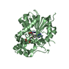

 PDBj
PDBj Assembly

 Mass: 96.063 Da / Num. of mol.: 3 / Source method: obtained synthetically / Formula: SO4
 Mass: 18.015 Da / Num. of mol.: 91 / Source method: isolated from a natural source / Formula: H2O
 Sample preparation
 Processing