 Movie
Movie Controller


+ Open data

- Basic information
| Entry | Database: PDB / ID: 1vif | ||||||
|---|---|---|---|---|---|---|---|
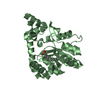
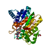
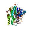
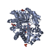
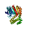
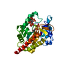
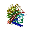
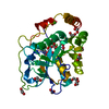
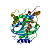
| Title | STRUCTURE OF DIHYDROFOLATE REDUCTASE | ||||||
 Components Components | DIHYDROFOLATE REDUCTASE | ||||||
 Keywords Keywords | OXIDOREDUCTASE / NADP / TRIMETHOPRIM RESISTANCE METHOTREXATE RESISTANCE / ONE-CARBON METABOLISM / PLASMID | ||||||
| Function / homology |  Function and homology information Function and homology informationresponse to methotrexate / dihydrofolate reductase / dihydrofolate reductase activity / tetrahydrofolate biosynthetic process / one-carbon metabolic process / response to xenobiotic stimulus / response to antibiotic Similarity search - Function | ||||||
| Biological species |  | ||||||
| Method |  X-RAY DIFFRACTION / ISOMORPHOUS REPLACEMENT / Resolution: 1.8 Å X-RAY DIFFRACTION / ISOMORPHOUS REPLACEMENT / Resolution: 1.8 Å | ||||||
 Authors Authors | Narayana, N. / Matthews, D.A. / Howell, E.E. / Xuong, N.-H. | ||||||
 Citation Citation | Journal: Nat.Struct.Biol. / Year: 1995 Title: A plasmid-encoded dihydrofolate reductase from trimethoprim-resistant bacteria has a novel D2-symmetric active site. Authors: Narayana, N. / Matthews, D.A. / Howell, E.E. / Nguyen-huu, X. #1: Journal: Adv.Exp.Med.Biol. / Year: 1993Title: Does R67 Dihydrofolate Reductase Possess a Proton Donor? Authors: Holland, J.C. / Linn, C.E. / Digiammarino, E. / Nichols, R. / Howell, E.E. #2: Journal: Biochemistry / Year: 1991Title: Construction of a Synthetic Gene for an R-Plasmid-Encoded Dihydrofolate Reductase and Studies on the Role of the N-Terminus in the Protein Authors: Reece, L.J. / Nichols, R. / Ogden, R.C. / Howell, E.E. #3: Journal: Biochemistry / Year: 1986Title: Crystal Structure of a Novel Trimethoprim-Resistant Dihydrofolate Reductase Specified in Escherichia Coli by R-Plasmid R67 Authors: Matthews, D.A. / Smith, S.L. / Baccanari, D.P. / Burchall, J.J. / Oatley, S.J. / Kraut, J. #4: Journal: J.Biol.Chem. / Year: 1979Title: The Amino Acid Sequence of the Trimethoprim-Resistant Dihydrofolate Reductase Specified in Escherichia Coli by R-Plasmid R67 Authors: Stone, D. / Smith, S.L. #5: Journal: Br.Med.J. / Year: 1972Title: Trimethoprim Resistance Determined by R Factors Authors: Fleming, M.P. / Datta, N. / Gruneberg, R.N. | ||||||
| History |
|
- Structure visualization
| Structure viewer | Molecule: MolmilJmol/JSmol |
|---|
- Downloads & links
-Download
| PDBx/mmCIF format | 1vif.cif.gz | 27.7 KB | Display | PDBx/mmCIF format |
|---|---|---|---|---|
| PDB format | pdb1vif.ent.gz | 17.2 KB | Display | PDB format |
| PDBx/mmJSON format | 1vif.json.gz | Tree view | PDBx/mmJSON format | |
| Others |  Other downloads Other downloads |
-Validation report
| Arichive directory | https://data.pdbj.org/pub/pdb/validation_reports/vi/1vifftp://data.pdbj.org/pub/pdb/validation_reports/vi/1vif | HTTPS FTP |
|---|
-Related structure data
| Related structure data |  1vieSC S: Starting model for refinement C: citing same article ( |
|---|---|
| Similar structure data |

-Links
 PDBj
PDBj


- Assembly
| Deposited unit | 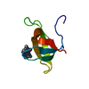
| ||||||||
|---|---|---|---|---|---|---|---|---|---|
| 1 | 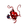
| ||||||||
| Unit cell |
|
-Components
| #1: Protein | Mass: 6732.528 Da / Num. of mol.: 1 Source method: isolated from a genetically manipulated source Source: (gene. exp.) Strain: TMP-RESISTANT, CONTAINING R67 DHFR OVERPRODUCING PLASMID PLZ1 Gene: SYNTHETIC GENE / Plasmid: PLZ1 / Production host: |
|---|---|
| #2: Chemical | ChemComp-FOL /   Mass: 441.397 Da / Num. of mol.: 1 / Source method: obtained synthetically / Formula: C19H19N7O6 Mass: 441.397 Da / Num. of mol.: 1 / Source method: obtained synthetically / Formula: C19H19N7O6 |
| #3: Water | ChemComp-HOH /  Mass: 18.015 Da / Num. of mol.: 44 / Source method: isolated from a natural source / Formula: H2O Mass: 18.015 Da / Num. of mol.: 44 / Source method: isolated from a natural source / Formula: H2O |
| Compound details | R67 PLASMID-ENCODED DHFR HAS 78 AMINO ACID RESIDUES. THE PRESENT STUDY DESCRIBES THE TRUNCATED FORM ...R67 PLASMID-ENCODED DHFR HAS 78 AMINO ACID RESIDUES. THE PRESENT STUDY DESCRIBES THE TRUNCATED FORM OF R67 DHFR (62 RESIDUES) OBTAINED BY CLEAVING THE FULL-LENGTH PROTEIN AT PHE 16 USING CHYMOTRYPS |
| Nonpolymer details | THE TWO MUTUALLY EXCLUSIVE FOLATE MOLECULES AT 1/4 OCCUPANCY ARE LABELLED AS FOL 1 WITH ALTERNATE ...THE TWO MUTUALLY EXCLUSIVE FOLATE MOLECULES AT 1/4 OCCUPANCY ARE LABELLED AS FOL 1 WITH ALTERNATE LOCATIONS A AND B. HOWEVER, DENSITY IS SEEN ONLY FOR THE PTERIDINE PORTION. THUS ATOMIC COORDINATE |
-Experimental details
-Experiment
| Experiment | Method: X-RAY DIFFRACTION / Number of used crystals: 1 |
|---|
- Sample preparation
| Crystal | Density Matthews: 2 Å3/Da / Density % sol: 40 % | |||||||||||||||||||||||||||||||||||
|---|---|---|---|---|---|---|---|---|---|---|---|---|---|---|---|---|---|---|---|---|---|---|---|---|---|---|---|---|---|---|---|---|---|---|---|---|
| Crystal grow | Method: vapor diffusion, hanging drop / pH: 8 Details: CRYSTALS WERE GROWN FROM HANGING- DROPS CONTAINING PROTEIN AT A FINAL CONCENTRATION OF ABOUT 18 MG/ML, 30 MM FOLATE, 40 MM BICINE BUFFER AT PH 8.0 AND 18% 2-METHYL-2,4-PENTANE DIOL (MPD). ...Details: CRYSTALS WERE GROWN FROM HANGING- DROPS CONTAINING PROTEIN AT A FINAL CONCENTRATION OF ABOUT 18 MG/ML, 30 MM FOLATE, 40 MM BICINE BUFFER AT PH 8.0 AND 18% 2-METHYL-2,4-PENTANE DIOL (MPD). DROPS WERE EQUILIBRATED AGAINST A RESERVOIR CONTAINING 100 MM KH2PO4 BUFFER AT PH 6.8 AND 50% MPD. THE CRYSTALS WERE FURTHER SOAKED IN 100 MM FOLATE FOR 3 DAYS., vapor diffusion - hanging drop PH range: 6.8-8.0 | |||||||||||||||||||||||||||||||||||
| Crystal grow | *PLUS Temperature: 4 ℃ / Method: vapor diffusion, hanging drop | |||||||||||||||||||||||||||||||||||
| Components of the solutions | *PLUS
|
-Data collection
| Diffraction | Mean temperature: 277 K |
|---|---|
| Diffraction source | Source: ROTATING ANODE / Type: RIGAKU RUH2R / Wavelength: 1.5418 |
| Detector | Type: XUONG-HAMLIN MULTIWIRE / Detector: AREA DETECTOR / Date: Mar 15, 1992 |
| Radiation | Monochromator: GRAPHITE(002) / Monochromatic (M) / Laue (L): M / Scattering type: x-ray |
| Radiation wavelength | Wavelength: 1.5418 Å / Relative weight: 1 |
| Reflection | Resolution: 1.8→10 Å / Num. obs: 6094 / % possible obs: 100 % / Observed criterion σ(I): 2 / Redundancy: 12 % / Biso Wilson estimate: 10.7 Å2 / Rsym value: 0.055 / Net I/σ(I): 12 |
| Reflection shell | Resolution: 1.8→1.86 Å / Redundancy: 8 % / Mean I/σ(I) obs: 2.2 / Rsym value: 0.16 / % possible all: 100 |
| Reflection | *PLUS Rmerge(I) obs: 0.055 |
- Processing
| Software |
| ||||||||||||||||||||||||||||||||||||||||||||||||||||||||||||
|---|---|---|---|---|---|---|---|---|---|---|---|---|---|---|---|---|---|---|---|---|---|---|---|---|---|---|---|---|---|---|---|---|---|---|---|---|---|---|---|---|---|---|---|---|---|---|---|---|---|---|---|---|---|---|---|---|---|---|---|---|---|
| Refinement | Method to determine structure: ISOMORPHOUS REPLACEMENT Starting model: PDB ENTRY 1VIE Resolution: 1.8→10 Å / σ(F): 1
| ||||||||||||||||||||||||||||||||||||||||||||||||||||||||||||
| Refine analyze | Luzzati coordinate error obs: 0.25 Å / Luzzati d res low obs: 10 Å | ||||||||||||||||||||||||||||||||||||||||||||||||||||||||||||
| Refinement step | Cycle: LAST / Resolution: 1.8→10 Å
| ||||||||||||||||||||||||||||||||||||||||||||||||||||||||||||
| Refine LS restraints |
| ||||||||||||||||||||||||||||||||||||||||||||||||||||||||||||
| Software | *PLUS Name: X-PLOR / Classification: refinement | ||||||||||||||||||||||||||||||||||||||||||||||||||||||||||||
| Refine LS restraints | *PLUS
|
