 Movie
Movie Controller

[English] 日本語
 Yorodumi
- PDB-1qtr: CRYSTAL STRUCTURE ANALYSIS OF THE PROLYL AMINOPEPTIDASE FROM SERR... -
+ Open data

- Basic information
| Entry | Database: PDB / ID: 1qtr | ||||||
|---|---|---|---|---|---|---|---|
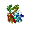
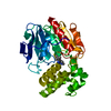
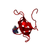
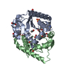
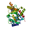
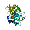
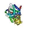
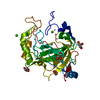
| Title | CRYSTAL STRUCTURE ANALYSIS OF THE PROLYL AMINOPEPTIDASE FROM SERRATIA MARCESCENS | ||||||
 Components Components | PROLYL AMINOPEPTIDASE | ||||||
 Keywords Keywords | HYDROLASE / ALPHA BETA HYDROLASE FOLD / PROLINE / PROLYL AMINOPEPTIDASE / SERRATIA / IMINOPEPTIDASE | ||||||
| Function / homology |  Function and homology information Function and homology informationprolyl aminopeptidase / aminopeptidase activity / proteolysis / cytoplasm Similarity search - Function | ||||||
| Biological species |  Serratia marcescens (bacteria) Serratia marcescens (bacteria) | ||||||
| Method |  X-RAY DIFFRACTION / Resolution: 2.32 Å X-RAY DIFFRACTION / Resolution: 2.32 Å | ||||||
 Authors Authors | Yoshimoto, T. / Kabashima, T. / Uchikawa, K. / Inoue, T. / Tanaka, N. | ||||||
 Citation Citation | Journal: J.Biochem.(Tokyo) / Year: 1999 Title: Crystal structure of prolyl aminopeptidase from Serratia marcescens. Authors: Yoshimoto, T. / Kabashima, T. / Uchikawa, K. / Inoue, T. / Tanaka, N. / Nakamura, K.T. / Tsuru, M. / Ito, K. #1: Journal: J.Biochem.(Tokyo) / Year: 1997Title: Prolyl Aminopeptidase from Serratia marcescens : Sequencing and Expression Authors: Kabashima, T. / Kitazono, A. / Kitano, A. / Inoue, K. / Yoshimoto, T. #2: Journal: J.Biochem.(Tokyo) / Year: 1994Title: Prolyl aminopeptidase is not a sulfhydryl enzyme: Identification of the active serine residue by site-directed mutagenesis Authors: Kitazono, A. / Ito, K. / Yoshimoto, T. | ||||||
| History |
|
- Structure visualization
| Structure viewer | Molecule: MolmilJmol/JSmol |
|---|
- Downloads & links
-Download
| PDBx/mmCIF format | 1qtr.cif.gz | 71.9 KB | Display | PDBx/mmCIF format |
|---|---|---|---|---|
| PDB format | pdb1qtr.ent.gz | 54.7 KB | Display | PDB format |
| PDBx/mmJSON format | 1qtr.json.gz | Tree view | PDBx/mmJSON format | |
| Others |  Other downloads Other downloads |
-Validation report
| Arichive directory | https://data.pdbj.org/pub/pdb/validation_reports/qt/1qtrftp://data.pdbj.org/pub/pdb/validation_reports/qt/1qtr | HTTPS FTP |
|---|
-Related structure data
| Similar structure data |
|---|


-Links
 PDBj
PDBj


- Assembly
| Deposited unit | 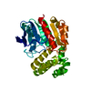
| ||||||||
|---|---|---|---|---|---|---|---|---|---|
| 1 |
| ||||||||
| Unit cell |
|
-Components
| #1: Protein | Mass: 36130.594 Da / Num. of mol.: 1 Source method: isolated from a genetically manipulated source Source: (gene. exp.) Serratia marcescens (bacteria) / Plasmid: PSPAP-HE / Production host: |
|---|
-Experimental details
-Experiment
| Experiment | Method: X-RAY DIFFRACTION / Number of used crystals: 1 |
|---|
- Sample preparation
| Crystal | Density Matthews: 2.5 Å3/Da / Density % sol: 50.79 % | |||||||||||||||||||||||||
|---|---|---|---|---|---|---|---|---|---|---|---|---|---|---|---|---|---|---|---|---|---|---|---|---|---|---|
| Crystal grow | Temperature: 297 K / Method: vapor diffusion, hanging drop / pH: 6.5 Details: PEG6000, SODIUM ACETATE, NA CACODYLATE/KH2PO4, pH 6.5, VAPOR DIFFUSION, HANGING DROP, temperature 297K | |||||||||||||||||||||||||
| Crystal grow | *PLUS Temperature: 20 ℃ / Details: Kabashima, T., (1997) J.Biochem.(Tokyo), 122, 601. | |||||||||||||||||||||||||
| Components of the solutions | *PLUS
|
-Data collection
| Diffraction | Mean temperature: 293 K |
|---|---|
| Diffraction source | Source: ROTATING ANODE / Type: RIGAKU RU200 / Wavelength: 1.5418 |
| Detector | Type: RIGAKU RAXIS IIC / Detector: IMAGE PLATE / Date: Oct 25, 1997 |
| Radiation | Protocol: SINGLE WAVELENGTH / Monochromatic (M) / Laue (L): M / Scattering type: x-ray |
| Radiation wavelength | Wavelength: 1.5418 Å / Relative weight: 1 |
| Reflection | Resolution: 2.32→10 Å / Num. obs: 45646 / % possible obs: 89.1 % / Rmerge(I) obs: 0.059 |
| Reflection | *PLUS Num. obs: 14917 / Num. measured all: 45646 |
- Processing
| Software |
| ||||||||||||||||
|---|---|---|---|---|---|---|---|---|---|---|---|---|---|---|---|---|---|
| Refinement | Resolution: 2.32→8 Å / σ(F): 1
| ||||||||||||||||
| Refinement step | Cycle: LAST / Resolution: 2.32→8 Å
| ||||||||||||||||
| Software | *PLUS Name: X-PLOR / Classification: refinement | ||||||||||||||||
| Refine LS restraints | *PLUS
|
