 Movie
Movie Controller

[English] 日本語
 Yorodumi
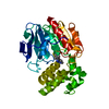
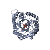
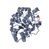
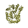
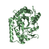
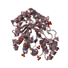
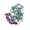
- PDB-4wlz: Crystal structure of mouse Xyloside xylosyltransferase 1 complexe... -
+ Open data

- Basic information
| Entry | Database: PDB / ID: 4wlz | ||||||
|---|---|---|---|---|---|---|---|
| Title | Crystal structure of mouse Xyloside xylosyltransferase 1 complexed with manganese and UDP | ||||||
 Components Components | Xyloside xylosyltransferase 1 | ||||||
 Keywords Keywords | TRANSFERASE / glycosyltransferase | ||||||
| Function / homology |  Function and homology information Function and homology informationxylosyl alpha-1,3-xylosyltransferase / xylosyl alpha-1,3-xylosyltransferase activity / protein O-linked glycosylation via glucose / UDP-xylosyltransferase activity / protein O-linked glycosylation via N-acetylgalactosamine / manganese ion binding / endoplasmic reticulum membrane / magnesium ion binding Similarity search - Function | ||||||
| Biological species |  | ||||||
| Method |  X-RAY DIFFRACTION / SYNCHROTRON / Resolution: 3.03 Å X-RAY DIFFRACTION / SYNCHROTRON / Resolution: 3.03 Å | ||||||
 Authors Authors | Yu, H. / Li, H. | ||||||
 Citation Citation | Journal: Nat.Chem.Biol. / Year: 2015 Title: Notch-modifying xylosyltransferase structures support an SNi-like retaining mechanism. Authors: Yu, H. / Takeuchi, M. / LeBarron, J. / Kantharia, J. / London, E. / Bakker, H. / Haltiwanger, R.S. / Li, H. / Takeuchi, H. | ||||||
| History |
|
- Structure visualization
| Structure viewer | Molecule: MolmilJmol/JSmol |
|---|
- Downloads & links
-Download
| PDBx/mmCIF format | 4wlz.cif.gz | 131.3 KB | Display | PDBx/mmCIF format |
|---|---|---|---|---|
| PDB format | pdb4wlz.ent.gz | 101.8 KB | Display | PDB format |
| PDBx/mmJSON format | 4wlz.json.gz | Tree view | PDBx/mmJSON format | |
| Others |  Other downloads Other downloads |
-Validation report
| Arichive directory | https://data.pdbj.org/pub/pdb/validation_reports/wl/4wlzftp://data.pdbj.org/pub/pdb/validation_reports/wl/4wlz | HTTPS FTP |
|---|
-Related structure data
| Related structure data |  4wlgC  4wlmC  4wm0C  4wmaC  4wmbC 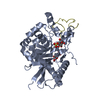 4wmiC  4wmkC  4wn2C C: citing same article ( |
|---|---|
| Similar structure data |


-Links
 PDBj
PDBj


- Assembly
| Deposited unit | 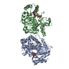
| ||||||||
|---|---|---|---|---|---|---|---|---|---|
| 1 | 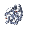
| ||||||||
| 2 | 
| ||||||||
| 3 | 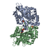
| ||||||||
| Unit cell |
|
-Components
| #1: Protein | Mass: 35295.500 Da / Num. of mol.: 2 Source method: isolated from a genetically manipulated source Source: (gene. exp.)  Homo sapiens (human) Homo sapiens (human)References: UniProt: Q3U4G3, Transferases; Glycosyltransferases; Pentosyltransferases #2: Chemical |   Mass: 54.938 Da / Num. of mol.: 2 / Source method: obtained synthetically / Formula: Mn Mass: 54.938 Da / Num. of mol.: 2 / Source method: obtained synthetically / Formula: Mn#3: Chemical |   Type: RNA linking / Mass: 404.161 Da / Num. of mol.: 2 / Source method: obtained synthetically / Formula: C9H14N2O12P2 / Comment: UDP*YM Type: RNA linking / Mass: 404.161 Da / Num. of mol.: 2 / Source method: obtained synthetically / Formula: C9H14N2O12P2 / Comment: UDP*YM#4: Chemical | ChemComp-SO4 / |   Mass: 96.063 Da / Num. of mol.: 1 / Source method: obtained synthetically / Formula: SO4 Mass: 96.063 Da / Num. of mol.: 1 / Source method: obtained synthetically / Formula: SO4Has protein modification | Y | |
|---|
-Experimental details
-Experiment
| Experiment | Method: X-RAY DIFFRACTION |
|---|
- Sample preparation
| Crystal | Density Matthews: 2.55 Å3/Da / Density % sol: 51.72 % |
|---|---|
| Crystal grow | Temperature: 293 K / Method: vapor diffusion, hanging drop / pH: 7.5 / Details: 20 mM HEPES, 1.5 M Li2SO4 |
-Data collection
| Diffraction | Mean temperature: 100 K |
|---|---|
| Diffraction source | Source: SYNCHROTRON / Site: NSLS  / Beamline: X25 / Wavelength: 1.1 Å / Beamline: X25 / Wavelength: 1.1 Å |
| Detector | Type: DECTRIS PILATUS 6M / Detector: PIXEL / Date: Apr 17, 2014 |
| Radiation | Protocol: SINGLE WAVELENGTH / Monochromatic (M) / Laue (L): M / Scattering type: x-ray |
| Radiation wavelength | Wavelength: 1.1 Å / Relative weight: 1 |
| Reflection | Resolution: 3.03→50 Å / Num. obs: 14615 / % possible obs: 99.3 % / Redundancy: 9 % / Rmerge(I) obs: 0.13 / Net I/σ(I): 13.7 |
| Reflection shell | Resolution: 3.03→3.14 Å / Redundancy: 6.8 % / Rmerge(I) obs: 0.547 / Mean I/σ(I) obs: 2.7 / % possible all: 95 |
- Processing
| Software | Name: REFMAC / Version: 5.6.0117 / Classification: refinement | ||||||||||||||||||||||||||||||||||||||||||||||||||||||||||||||||||||||||||||||||||||||||||||||||||||||||||||||||||||||||||||||||||||||||||||||||||||||||||||||||||||||||||||||||||||||
|---|---|---|---|---|---|---|---|---|---|---|---|---|---|---|---|---|---|---|---|---|---|---|---|---|---|---|---|---|---|---|---|---|---|---|---|---|---|---|---|---|---|---|---|---|---|---|---|---|---|---|---|---|---|---|---|---|---|---|---|---|---|---|---|---|---|---|---|---|---|---|---|---|---|---|---|---|---|---|---|---|---|---|---|---|---|---|---|---|---|---|---|---|---|---|---|---|---|---|---|---|---|---|---|---|---|---|---|---|---|---|---|---|---|---|---|---|---|---|---|---|---|---|---|---|---|---|---|---|---|---|---|---|---|---|---|---|---|---|---|---|---|---|---|---|---|---|---|---|---|---|---|---|---|---|---|---|---|---|---|---|---|---|---|---|---|---|---|---|---|---|---|---|---|---|---|---|---|---|---|---|---|---|---|
| Refinement | Resolution: 3.03→50 Å / Cor.coef. Fo:Fc: 0.914 / Cor.coef. Fo:Fc free: 0.862 / Cross valid method: THROUGHOUT / ESU R Free: 0.525 / Stereochemistry target values: MAXIMUM LIKELIHOOD / Details: HYDROGENS HAVE BEEN USED IF PRESENT IN THE INPUT
| ||||||||||||||||||||||||||||||||||||||||||||||||||||||||||||||||||||||||||||||||||||||||||||||||||||||||||||||||||||||||||||||||||||||||||||||||||||||||||||||||||||||||||||||||||||||
| Solvent computation | Ion probe radii: 0.8 Å / Shrinkage radii: 0.8 Å / VDW probe radii: 1.2 Å / Solvent model: MASK | ||||||||||||||||||||||||||||||||||||||||||||||||||||||||||||||||||||||||||||||||||||||||||||||||||||||||||||||||||||||||||||||||||||||||||||||||||||||||||||||||||||||||||||||||||||||
| Displacement parameters | Biso mean: 58.433 Å2
| ||||||||||||||||||||||||||||||||||||||||||||||||||||||||||||||||||||||||||||||||||||||||||||||||||||||||||||||||||||||||||||||||||||||||||||||||||||||||||||||||||||||||||||||||||||||
| Refinement step | Cycle: 1 / Resolution: 3.03→50 Å
| ||||||||||||||||||||||||||||||||||||||||||||||||||||||||||||||||||||||||||||||||||||||||||||||||||||||||||||||||||||||||||||||||||||||||||||||||||||||||||||||||||||||||||||||||||||||
| Refine LS restraints |
|