 Movie
Movie Controller

[English] 日本語
 Yorodumi
- PDB-1x2e: The crystal structure of prolyl aminopeptidase complexed with Ala... -
+ Open data

- Basic information
| Entry | Database: PDB / ID: 1x2e | ||||||
|---|---|---|---|---|---|---|---|
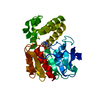
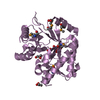
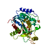
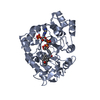
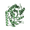
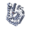
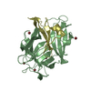
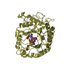
| Title | The crystal structure of prolyl aminopeptidase complexed with Ala-TBODA | ||||||
 Components Components | Proline iminopeptidase | ||||||
 Keywords Keywords | HYDROLASE / prolyl aminopeptidase / prolyl iminopeptidase / binary complex / alpha/beta-hydrolase fold | ||||||
| Function / homology |  Function and homology information Function and homology informationprolyl aminopeptidase / aminopeptidase activity / proteolysis / cytoplasm Similarity search - Function | ||||||
| Biological species |  Serratia marcescens (bacteria) Serratia marcescens (bacteria) | ||||||
| Method |  X-RAY DIFFRACTION / SYNCHROTRON / FOURIER SYNTHESIS / Resolution: 2.4 Å X-RAY DIFFRACTION / SYNCHROTRON / FOURIER SYNTHESIS / Resolution: 2.4 Å | ||||||
 Authors Authors | Nakajima, Y. / Ito, K. / Sakata, M. / Xu, Y. / Matsubara, F. / Hatakeyama, S. / Yoshimoto, T. | ||||||
 Citation Citation | Journal: J.Bacteriol. / Year: 2006 Title: Unusual extra space at the active site and high activity for acetylated hydroxyproline of prolyl aminopeptidase from Serratia marcescens Authors: Nakajima, Y. / Ito, K. / Sakata, M. / Xu, Y. / Nakashima, K. / Matsubara, F. / Hatakeyama, S. / Yoshimoto, T. | ||||||
| History |
|
- Structure visualization
| Structure viewer | Molecule: MolmilJmol/JSmol |
|---|
- Downloads & links
-Download
| PDBx/mmCIF format | 1x2e.cif.gz | 76.7 KB | Display | PDBx/mmCIF format |
|---|---|---|---|---|
| PDB format | pdb1x2e.ent.gz | 56.2 KB | Display | PDB format |
| PDBx/mmJSON format | 1x2e.json.gz | Tree view | PDBx/mmJSON format | |
| Others |  Other downloads Other downloads |
-Validation report
| Arichive directory | https://data.pdbj.org/pub/pdb/validation_reports/x2/1x2eftp://data.pdbj.org/pub/pdb/validation_reports/x2/1x2e | HTTPS FTP |
|---|
-Related structure data
| Related structure data | 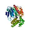 1x2bC  1qtrS S: Starting model for refinement C: citing same article ( |
|---|---|
| Similar structure data |


-Links
 PDBj
PDBj


- Assembly
| Deposited unit | 
| ||||||||
|---|---|---|---|---|---|---|---|---|---|
| 1 |
| ||||||||
| Unit cell |
| ||||||||
| Details | The biological assembly is the monomer in asymmetric unit. |
-Components
| #1: Protein | Mass: 36130.594 Da / Num. of mol.: 1 Source method: isolated from a genetically manipulated source Source: (gene. exp.) Serratia marcescens (bacteria) / Plasmid: pUC19 / Production host: |
|---|---|
| #2: Chemical | ChemComp-ATX / (  Mass: 197.234 Da / Num. of mol.: 1 / Source method: obtained synthetically / Formula: C9H15N3O2 Mass: 197.234 Da / Num. of mol.: 1 / Source method: obtained synthetically / Formula: C9H15N3O2 |
| #3: Water | ChemComp-HOH /  Mass: 18.015 Da / Num. of mol.: 75 / Source method: isolated from a natural source / Formula: H2O Mass: 18.015 Da / Num. of mol.: 75 / Source method: isolated from a natural source / Formula: H2O |
-Experimental details
-Experiment
| Experiment | Method: X-RAY DIFFRACTION / Number of used crystals: 1 |
|---|
- Sample preparation
| Crystal | Density Matthews: 2.49 Å3/Da / Density % sol: 50.6 % |
|---|---|
| Crystal grow | Temperature: 293 K / Method: vapor diffusion, hanging drop / pH: 6.5 Details: PEG6000, cacodylate, sodium acetate, pH 6.5, VAPOR DIFFUSION, HANGING DROP, temperature 293K |
-Data collection
| Diffraction | Mean temperature: 298 K |
|---|---|
| Diffraction source | Source: SYNCHROTRON / Site: Photon Factory  / Beamline: BL-18B / Wavelength: 1.15 Å / Beamline: BL-18B / Wavelength: 1.15 Å |
| Detector | Type: ADSC QUANTUM 4 / Detector: CCD / Date: Dec 2, 2004 |
| Radiation | Protocol: SINGLE WAVELENGTH / Monochromatic (M) / Laue (L): M / Scattering type: x-ray |
| Radiation wavelength | Wavelength: 1.15 Å / Relative weight: 1 |
| Reflection | Resolution: 2.4→50 Å / Num. all: 15059 / Num. obs: 15059 / % possible obs: 99.8 % / Observed criterion σ(F): 0 / Observed criterion σ(I): 0 / Redundancy: 7.7 % / Rmerge(I) obs: 0.066 / Net I/σ(I): 45.4 |
| Reflection shell | Resolution: 2.4→2.49 Å / Redundancy: 7.9 % / Rmerge(I) obs: 0.283 / Mean I/σ(I) obs: 10.4 / Num. unique all: 1467 / % possible all: 100 |
- Processing
| Software |
| |||||||||||||||||||||||||
|---|---|---|---|---|---|---|---|---|---|---|---|---|---|---|---|---|---|---|---|---|---|---|---|---|---|---|
| Refinement | Method to determine structure: FOURIER SYNTHESIS Starting model: 1QTR Resolution: 2.4→30 Å / Isotropic thermal model: anisotropic / Cross valid method: THROUGHOUT / σ(F): 0 / Stereochemistry target values: Engh & Huber
| |||||||||||||||||||||||||
| Displacement parameters | Biso mean: 35.3 Å2
| |||||||||||||||||||||||||
| Refine analyze |
| |||||||||||||||||||||||||
| Refinement step | Cycle: LAST / Resolution: 2.4→30 Å
| |||||||||||||||||||||||||
| Refine LS restraints |
| |||||||||||||||||||||||||
| LS refinement shell | Resolution: 2.4→2.49 Å
|
