 Movie
Movie Controller

[English] 日本語
 Yorodumi
- PDB-3azs: Diverse Substrates Recognition Mechanism Revealed by Thermotoga m... -
+ Open data

- Basic information
| Entry | Database: PDB / ID: 3azs | |||||||||
|---|---|---|---|---|---|---|---|---|---|---|
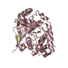
| Title | Diverse Substrates Recognition Mechanism Revealed by Thermotoga maritima Cel5A Structures in Complex with Mannotriose | |||||||||
 Components Components | Endoglucanase | |||||||||
 Keywords Keywords | HYDROLASE / cellulose / cellulase / biofuel / Tim Barrel | |||||||||
| Function / homology |  Function and homology information Function and homology information | |||||||||
| Biological species |   Thermotoga maritima (bacteria) Thermotoga maritima (bacteria) | |||||||||
| Method |  X-RAY DIFFRACTION / SYNCHROTRON / MOLECULAR REPLACEMENT / Resolution: 1.69 Å X-RAY DIFFRACTION / SYNCHROTRON / MOLECULAR REPLACEMENT / Resolution: 1.69 Å | |||||||||
 Authors Authors | Wu, T.H. / Huang, C.H. / Ko, T.P. / Lai, H.L. / Ma, Y. / Chen, C.C. / Cheng, Y.S. / Liu, J.R. / Guo, R.T. | |||||||||
 Citation Citation | Journal: Biochim.Biophys.Acta / Year: 2011 Title: Diverse substrate recognition mechanism revealed by Thermotoga maritima Cel5A structures in complex with cellotetraose, cellobiose and mannotriose Authors: Wu, T.H. / Huang, C.H. / Ko, T.P. / Lai, H.L. / Ma, Y. / Chen, C.C. / Cheng, Y.S. / Liu, J.R. / Guo, R.T. | |||||||||
| History |
|
- Structure visualization
| Structure viewer | Molecule: MolmilJmol/JSmol |
|---|
- Downloads & links
-Download
| PDBx/mmCIF format | 3azs.cif.gz | 155.4 KB | Display | PDBx/mmCIF format |
|---|---|---|---|---|
| PDB format | pdb3azs.ent.gz | 121.3 KB | Display | PDB format |
| PDBx/mmJSON format | 3azs.json.gz | Tree view | PDBx/mmJSON format | |
| Others |  Other downloads Other downloads |
-Validation report
| Arichive directory | https://data.pdbj.org/pub/pdb/validation_reports/az/3azsftp://data.pdbj.org/pub/pdb/validation_reports/az/3azs | HTTPS FTP |
|---|
-Related structure data
| Related structure data |  3amcSC  3amdC  3amgC  3aofC  3azrC  3aztC S: Starting model for refinement C: citing same article ( |
|---|---|
| Similar structure data |


-Links
 PDBj
PDBj- Assembly
| Deposited unit | 
| ||||||||
|---|---|---|---|---|---|---|---|---|---|
| 1 | 
| ||||||||
| 2 | 
| ||||||||
| Unit cell |
|
-Components
| #1: Protein | Mass: 37380.488 Da / Num. of mol.: 2 / Mutation: E253A Source method: isolated from a genetically manipulated source Source: (gene. exp.) Thermotoga maritima (bacteria) / Strain: MSB8 / Gene: TM_1751 / Plasmid: pET32 Xa/LIC / Production host: #2: Polysaccharide | Source method: isolated from a genetically manipulated source #3: Water | ChemComp-HOH / |  Mass: 18.015 Da / Num. of mol.: 674 / Source method: isolated from a natural source / Formula: H2O Mass: 18.015 Da / Num. of mol.: 674 / Source method: isolated from a natural source / Formula: H2O |
|---|
-Experimental details
-Experiment
| Experiment | Method: X-RAY DIFFRACTION / Number of used crystals: 1 |
|---|
- Sample preparation
| Crystal | Density Matthews: 2.04 Å3/Da / Density % sol: 39.83 % |
|---|---|
| Crystal grow | Temperature: 293 K / Method: vapor diffusion, hanging drop / pH: 8.5 Details: 0.1M tris pH8.5 , 0.4M NaCl, 28% PEG 3350, VAPOR DIFFUSION, HANGING DROP, temperature 293K |
-Data collection
| Diffraction | Mean temperature: 100 K |
|---|---|
| Diffraction source | Source: SYNCHROTRON / Site: NSRRC  / Beamline: BL13C1 / Wavelength: 1 Å / Beamline: BL13C1 / Wavelength: 1 Å |
| Detector | Type: ADSC QUANTUM 315r / Detector: CCD / Date: Apr 20, 2011 |
| Radiation | Protocol: SINGLE WAVELENGTH / Monochromatic (M) / Laue (L): M / Scattering type: x-ray |
| Radiation wavelength | Wavelength: 1 Å / Relative weight: 1 |
| Reflection | Resolution: 1.69→25 Å / Num. obs: 67472 / % possible obs: 99.4 % / Observed criterion σ(I): 2 / Redundancy: 4.1 % / Rsym value: 0.043 / Net I/σ(I): 30.1 |
| Reflection shell | Resolution: 1.69→1.75 Å / Redundancy: 4 % / Mean I/σ(I) obs: 10.6 / Num. unique all: 6608 / Rsym value: 0.124 / % possible all: 97.4 |
- Processing
| Software |
| ||||||||||||||||||||
|---|---|---|---|---|---|---|---|---|---|---|---|---|---|---|---|---|---|---|---|---|---|
| Refinement | Method to determine structure: MOLECULAR REPLACEMENT Starting model: PDB ENTRY 3AMC Resolution: 1.69→25 Å / σ(F): 2 / Stereochemistry target values: ML
| ||||||||||||||||||||
| Solvent computation | Bsol: 44.6583 Å2 | ||||||||||||||||||||
| Displacement parameters | Biso mean: 15.2534 Å2
| ||||||||||||||||||||
| Refinement step | Cycle: LAST / Resolution: 1.69→25 Å
| ||||||||||||||||||||
| Refine LS restraints |
| ||||||||||||||||||||
| Xplor file |
|
