 Movie
Movie Controller

[English] 日本語
 Yorodumi
- PDB-3amd: Crystal structures of Thermotoga maritima Cel5A, apo form and tet... -
+ Open data

- Basic information
| Entry | Database: PDB / ID: 3amd | ||||||
|---|---|---|---|---|---|---|---|
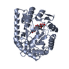
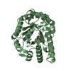
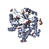
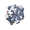
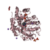
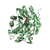
| Title | Crystal structures of Thermotoga maritima Cel5A, apo form and tetramer/au | ||||||
 Components Components | Endoglucanase | ||||||
 Keywords Keywords | HYDROLASE / glycosyl hydrolase family 5 / cellulase / biofuel / hyperthermostable / TIM-barrel | ||||||
| Function / homology |  Function and homology information Function and homology information | ||||||
| Biological species |   Thermotoga maritima (bacteria) Thermotoga maritima (bacteria) | ||||||
| Method |  X-RAY DIFFRACTION / SYNCHROTRON / MOLECULAR REPLACEMENT / Resolution: 2 Å X-RAY DIFFRACTION / SYNCHROTRON / MOLECULAR REPLACEMENT / Resolution: 2 Å | ||||||
 Authors Authors | Wu, T.H. / Huang, C.H. / Ko, T.P. / Lai, H.L. / Ma, Y. / Cheng, Y.S. / Liu, J.R. / Guo, R.T. | ||||||
 Citation Citation | Journal: Biochim.Biophys.Acta / Year: 2011 Title: Diverse substrate recognition mechanism revealed by Thermotoga maritima Cel5A structures in complex with cellotetraose, cellobiose and mannotriose Authors: Wu, T.H. / Huang, C.H. / Ko, T.P. / Lai, H.L. / Ma, Y. / Chen, C.C. / Cheng, Y.S. / Liu, J.R. / Guo, R.T. | ||||||
| History |
|
- Structure visualization
| Structure viewer | Molecule: MolmilJmol/JSmol |
|---|
- Downloads & links
-Download
| PDBx/mmCIF format | 3amd.cif.gz | 273.5 KB | Display | PDBx/mmCIF format |
|---|---|---|---|---|
| PDB format | pdb3amd.ent.gz | 222.5 KB | Display | PDB format |
| PDBx/mmJSON format | 3amd.json.gz | Tree view | PDBx/mmJSON format | |
| Others |  Other downloads Other downloads |
-Validation report
| Arichive directory | https://data.pdbj.org/pub/pdb/validation_reports/am/3amdftp://data.pdbj.org/pub/pdb/validation_reports/am/3amd | HTTPS FTP |
|---|
-Related structure data
| Related structure data |  3amcC 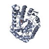 3amgC  3aofC  3azrC  3azsC  3aztC C: citing same article ( |
|---|---|
| Similar structure data |


-Links
 PDBj
PDBj- Assembly
| Deposited unit | 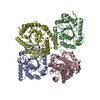
| ||||||||
|---|---|---|---|---|---|---|---|---|---|
| 1 | 
| ||||||||
| 2 | 
| ||||||||
| 3 | 
| ||||||||
| 4 | 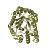
| ||||||||
| Unit cell |
|
-Components
| #1: Protein | Mass: 37438.523 Da / Num. of mol.: 4 Source method: isolated from a genetically manipulated source Source: (gene. exp.) Thermotoga maritima (bacteria) / Strain: MSB8 / Gene: TM_1751 / Plasmid: pET32 Xa/LIC / Production host: #2: Water | ChemComp-HOH / |  Mass: 18.015 Da / Num. of mol.: 761 / Source method: isolated from a natural source / Formula: H2O Mass: 18.015 Da / Num. of mol.: 761 / Source method: isolated from a natural source / Formula: H2O |
|---|
-Experimental details
-Experiment
| Experiment | Method: X-RAY DIFFRACTION / Number of used crystals: 1 |
|---|
- Sample preparation
| Crystal | Density Matthews: 1.94 Å3/Da / Density % sol: 36.47 % |
|---|---|
| Crystal grow | Temperature: 298 K / Method: vapor diffusion, sitting drop / pH: 8.5 Details: 0.2M NaCl, 0.1M Tris, 25% PEG 3350, pH 8.5, VAPOR DIFFUSION, SITTING DROP, temperature 298K |
-Data collection
| Diffraction | Mean temperature: 100 K |
|---|---|
| Diffraction source | Source: SYNCHROTRON / Site: NSRRC  / Beamline: BL13C1 / Wavelength: 1 Å / Beamline: BL13C1 / Wavelength: 1 Å |
| Detector | Type: ADSC QUANTUM 315r / Detector: CCD / Date: Dec 31, 2009 |
| Radiation | Protocol: SINGLE WAVELENGTH / Monochromatic (M) / Laue (L): M / Scattering type: x-ray |
| Radiation wavelength | Wavelength: 1 Å / Relative weight: 1 |
| Reflection | Resolution: 2→25 Å / Num. obs: 77633 / % possible obs: 99.7 % / Observed criterion σ(I): 0 / Redundancy: 4.8 % / Biso Wilson estimate: 9.8 Å2 / Rsym value: 0.085 / Net I/σ(I): 18.5 |
| Reflection shell | Resolution: 2→2.07 Å / Redundancy: 4.8 % / Mean I/σ(I) obs: 3.8 / Rsym value: 0.401 / % possible all: 99.9 |
- Processing
| Software |
| ||||||||||||||||||||||||||||||||||||||||||||||||||||||||||||||||||||||||||||||||
|---|---|---|---|---|---|---|---|---|---|---|---|---|---|---|---|---|---|---|---|---|---|---|---|---|---|---|---|---|---|---|---|---|---|---|---|---|---|---|---|---|---|---|---|---|---|---|---|---|---|---|---|---|---|---|---|---|---|---|---|---|---|---|---|---|---|---|---|---|---|---|---|---|---|---|---|---|---|---|---|---|---|
| Refinement | Method to determine structure: MOLECULAR REPLACEMENT / Resolution: 2→25 Å / Occupancy max: 1 / Occupancy min: 1 / Isotropic thermal model: RESTRAINED / Cross valid method: THROUGHOUT / σ(F): 0 / Details: BULK SOLVENT MODEL USED
| ||||||||||||||||||||||||||||||||||||||||||||||||||||||||||||||||||||||||||||||||
| Solvent computation | Solvent model: FLAT MODEL / Bsol: 33.5088 Å2 | ||||||||||||||||||||||||||||||||||||||||||||||||||||||||||||||||||||||||||||||||
| Displacement parameters | Biso mean: 19.9348 Å2
| ||||||||||||||||||||||||||||||||||||||||||||||||||||||||||||||||||||||||||||||||
| Refine analyze |
| ||||||||||||||||||||||||||||||||||||||||||||||||||||||||||||||||||||||||||||||||
| Refinement step | Cycle: LAST / Resolution: 2→25 Å
| ||||||||||||||||||||||||||||||||||||||||||||||||||||||||||||||||||||||||||||||||
| Refine LS restraints |
| ||||||||||||||||||||||||||||||||||||||||||||||||||||||||||||||||||||||||||||||||
| LS refinement shell | Resolution: 2→2.13 Å / Rfactor Rfree error: 0.013 / Total num. of bins used: 6
| ||||||||||||||||||||||||||||||||||||||||||||||||||||||||||||||||||||||||||||||||
| Xplor file |
|
