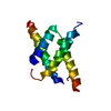
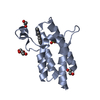
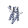
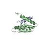
Entry Database : PDB / ID : 2p4eTitle Crystal Structure of PCSK9 Proprotein convertase subtilisin/kexin type 9 Keywords / / / / / Function / homology Function Domain/homology Component
/ / / / / / / / / / / / / / / / / / / / / / / / / / / / / / / / / / / / / / / / / / / / / / / / / / / / / / / / / / / / / / / / / / / / / / / / / / / / / / / / / / / / / / / / / / / / / / / / / / / / Biological species Homo sapiens (human)Method / / / Resolution : 1.98 Å Authors Cunningham, D. / Danley, D.E. / Geoghegan, F.K. / Griffor, M.C. / Hawkins, J.L. / Qiu, X. Journal : Nat.Struct.Mol.Biol. / Year : 2007Title : Structural and biophysical studies of PCSK9 and its mutants linked to familial hypercholesterolemia.Authors: Cunningham, D. / Danley, D.E. / Geoghegan, K.F. / Griffor, M.C. / Hawkins, J.L. / Subashi, T.A. / Varghese, A.H. / Ammirati, M.J. / Culp, J.S. / Hoth, L.R. / Mansour, M.N. / McGrath, K.M. / ... Authors : Cunningham, D. / Danley, D.E. / Geoghegan, K.F. / Griffor, M.C. / Hawkins, J.L. / Subashi, T.A. / Varghese, A.H. / Ammirati, M.J. / Culp, J.S. / Hoth, L.R. / Mansour, M.N. / McGrath, K.M. / Seddon, A.P. / Shenolikar, S. / Stutzman-Engwall, K.J. / Warren, L.C. / Xia, D. / Qiu, X. History Deposition Mar 12, 2007 Deposition site / Processing site Revision 1.0 Apr 10, 2007 Provider / Type Revision 1.1 May 1, 2008 Group Revision 1.2 Jul 13, 2011 Group / Version format complianceRevision 1.3 Aug 30, 2023 Group Data collection / Database references ... Data collection / Database references / Derived calculations / Refinement description Category chem_comp_atom / chem_comp_bond ... chem_comp_atom / chem_comp_bond / database_2 / pdbx_initial_refinement_model / pdbx_struct_conn_angle / struct_conn / struct_site Item _database_2.pdbx_DOI / _database_2.pdbx_database_accession ... _database_2.pdbx_DOI / _database_2.pdbx_database_accession / _pdbx_struct_conn_angle.ptnr1_auth_asym_id / _pdbx_struct_conn_angle.ptnr1_auth_comp_id / _pdbx_struct_conn_angle.ptnr1_auth_seq_id / _pdbx_struct_conn_angle.ptnr1_label_asym_id / _pdbx_struct_conn_angle.ptnr1_label_atom_id / _pdbx_struct_conn_angle.ptnr1_label_comp_id / _pdbx_struct_conn_angle.ptnr1_label_seq_id / _pdbx_struct_conn_angle.ptnr3_auth_asym_id / _pdbx_struct_conn_angle.ptnr3_auth_comp_id / _pdbx_struct_conn_angle.ptnr3_auth_seq_id / _pdbx_struct_conn_angle.ptnr3_label_asym_id / _pdbx_struct_conn_angle.ptnr3_label_atom_id / _pdbx_struct_conn_angle.ptnr3_label_comp_id / _pdbx_struct_conn_angle.ptnr3_label_seq_id / _pdbx_struct_conn_angle.value / _struct_conn.pdbx_dist_value / _struct_conn.ptnr1_auth_comp_id / _struct_conn.ptnr1_auth_seq_id / _struct_conn.ptnr1_label_asym_id / _struct_conn.ptnr1_label_atom_id / _struct_conn.ptnr1_label_comp_id / _struct_conn.ptnr1_label_seq_id / _struct_conn.ptnr2_auth_asym_id / _struct_conn.ptnr2_auth_comp_id / _struct_conn.ptnr2_auth_seq_id / _struct_conn.ptnr2_label_asym_id / _struct_conn.ptnr2_label_atom_id / _struct_conn.ptnr2_label_comp_id / _struct_conn.ptnr2_label_seq_id / _struct_site.pdbx_auth_asym_id / _struct_site.pdbx_auth_comp_id / _struct_site.pdbx_auth_seq_id Revision 1.4 Oct 30, 2024 Group / Category / pdbx_modification_feature
Show all Show less
 Movie
Movie Controller


 Open data
 Basic information
 Components
 Keywords
 Function and homology information
 Homo sapiens (human)
 X-RAY DIFFRACTION /
  Authors
 Citation
 Structure visualization
 Downloads & links
 Other downloads
 PDBj
PDBj


 Assembly


 Cricetulus griseus (Chinese hamster)
 Mass: 200.590 Da / Num. of mol.: 2 / Source method: obtained synthetically / Formula: Hg
 Mass: 18.015 Da / Num. of mol.: 371 / Source method: isolated from a natural source / Formula: H2O
 Sample preparation
 / Beamline: 17-ID / Wavelength: 1 Å
 Processing