 Movie
Movie Controller

[English] 日本語
 Yorodumi
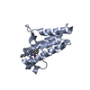
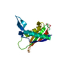
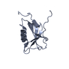
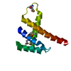
- PDB-1g1e: NMR STRUCTURE OF THE HUMAN MAD1 TRANSREPRESSION DOMAIN SID IN COM... -
+ Open data

- Basic information
| Entry | Database: PDB / ID: 1g1e | ||||||
|---|---|---|---|---|---|---|---|
| Title | NMR STRUCTURE OF THE HUMAN MAD1 TRANSREPRESSION DOMAIN SID IN COMPLEX WITH MAMMALIAN SIN3A PAH2 DOMAIN | ||||||
 Components Components |
| ||||||
 Keywords Keywords | TRANSCRIPTION / Four-helix bundle / Protein-peptide Complex | ||||||
| Function / homology |  Function and homology information Function and homology informationcellular response to tert-butyl hydroperoxide / Regulation of MECP2 expression and activity / Regulation of MITF-M-dependent genes involved in apoptosis / Mad-Max complex / response to methylglyoxal / Regulation of MITF-M-dependent genes involved in cell cycle and proliferation / SUMOylation of transcription cofactors / regulation of hormone levels / cerebral cortex neuron differentiation / RUNX1 regulates genes involved in megakaryocyte differentiation and platelet function ...cellular response to tert-butyl hydroperoxide / Regulation of MECP2 expression and activity / Regulation of MITF-M-dependent genes involved in apoptosis / Mad-Max complex / response to methylglyoxal / Regulation of MITF-M-dependent genes involved in cell cycle and proliferation / SUMOylation of transcription cofactors / regulation of hormone levels / cerebral cortex neuron differentiation / RUNX1 regulates genes involved in megakaryocyte differentiation and platelet function / Regulation of lipid metabolism by PPARalpha / negative regulation of circadian rhythm / Cytoprotection by HMOX1 / negative regulation of stem cell population maintenance / cellular response to dopamine / regulation of axon extension / negative regulation of protein localization to nucleus / type I interferon-mediated signaling pathway / Sin3-type complex / histone deacetylase complex / positive regulation of stem cell population maintenance / positive regulation of G2/M transition of mitotic cell cycle / hematopoietic progenitor cell differentiation / transcription regulator inhibitor activity / positive regulation of defense response to virus by host / transcription repressor complex / positive regulation of neuron differentiation / activation of innate immune response / negative regulation of cell migration / negative regulation of transforming growth factor beta receptor signaling pathway / cellular response to glucose stimulus / kinetochore / DNA-binding transcription repressor activity, RNA polymerase II-specific / transcription corepressor activity / intracellular protein localization / rhythmic process / heterochromatin formation / transcription regulator complex / in utero embryonic development / RNA polymerase II-specific DNA-binding transcription factor binding / DNA-binding transcription factor activity, RNA polymerase II-specific / DNA replication / protein dimerization activity / RNA polymerase II cis-regulatory region sequence-specific DNA binding / negative regulation of DNA-templated transcription / chromatin binding / regulation of transcription by RNA polymerase II / regulation of DNA-templated transcription / negative regulation of apoptotic process / chromatin / protein-containing complex binding / nucleolus / negative regulation of transcription by RNA polymerase II / protein-containing complex / mitochondrion / DNA binding / RNA binding / nucleoplasm / nucleus / cytosol Similarity search - Function | ||||||
| Biological species |  | ||||||
| Method | SOLUTION NMR / distance geometry, simulated annealing | ||||||
| Model type details | minimized average | ||||||
 Authors Authors | Brubaker, K. / Cowley, S.M. / Huang, K. / Eisenman, R.N. / Radhakrishnan, I. | ||||||
 Citation Citation | Journal: Cell(Cambridge,Mass.) / Year: 2000 Title: Solution structure of the interacting domains of the Mad-Sin3 complex: implications for recruitment of a chromatin-modifying complex. Authors: Brubaker, K. / Cowley, S.M. / Huang, K. / Loo, L. / Yochum, G.S. / Ayer, D.E. / Eisenman, R.N. / Radhakrishnan, I. | ||||||
| History |
|
- Structure visualization
| Structure viewer | Molecule: MolmilJmol/JSmol |
|---|
- Downloads & links
-Download
| PDBx/mmCIF format | 1g1e.cif.gz | 502.9 KB | Display | PDBx/mmCIF format |
|---|---|---|---|---|
| PDB format | pdb1g1e.ent.gz | 420.5 KB | Display | PDB format |
| PDBx/mmJSON format | 1g1e.json.gz | Tree view | PDBx/mmJSON format | |
| Others |  Other downloads Other downloads |
-Validation report
| Arichive directory | https://data.pdbj.org/pub/pdb/validation_reports/g1/1g1eftp://data.pdbj.org/pub/pdb/validation_reports/g1/1g1e | HTTPS FTP |
|---|
-Related structure data
| Similar structure data |
|---|


-Links
 PDBj
PDBj


- Assembly
| Deposited unit | 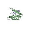
| |||||||||
|---|---|---|---|---|---|---|---|---|---|---|
| 1 |
| |||||||||
| NMR ensembles |
|
-Components
| #1: Protein/peptide | Mass: 1968.302 Da / Num. of mol.: 1 Fragment: SIN3 INTERACTION DOMAIN (SID) TRANSREPRESSION DOMAIN Source method: obtained synthetically Details: The peptide sequence was synthesized via automated methods. The sequence is naturally found in Homo sapiens (human). References: GenBank: 4505069, UniProt: Q05195*PLUS |
|---|---|
| #2: Protein | Mass: 10343.438 Da / Num. of mol.: 1 / Fragment: PAIRED AMPHIPATHIC HELIX 2 (PAH2 REPEAT) Source method: isolated from a genetically manipulated source Source: (gene. exp.)  |
-Experimental details
-Experiment
| Experiment | Method: SOLUTION NMR | ||||||||||||||||||||||||||||||||||||
|---|---|---|---|---|---|---|---|---|---|---|---|---|---|---|---|---|---|---|---|---|---|---|---|---|---|---|---|---|---|---|---|---|---|---|---|---|---|
| NMR experiment |
| ||||||||||||||||||||||||||||||||||||
| NMR details | Text: Interproton NOEs were assigned iteratively but manually during structure refinement. |
- Sample preparation
| Details |
| ||||||||||||
|---|---|---|---|---|---|---|---|---|---|---|---|---|---|
| Sample conditions | Ionic strength: 20 mM phosphate / pH: 6 / Pressure: ambient / Temperature: 300 K | ||||||||||||
| Crystal grow | *PLUS Method: other / Details: NMR |
-NMR measurement
| NMR spectrometer |
|
|---|
- Processing
| NMR software |
| ||||||||||||||||||||
|---|---|---|---|---|---|---|---|---|---|---|---|---|---|---|---|---|---|---|---|---|---|
| Refinement | Method: distance geometry, simulated annealing / Software ordinal: 1 Details: The structures are based on 1612 unique distance constraints, 198 torsion angle constraints and 72 JHNHA coupling constant constraints. | ||||||||||||||||||||
| NMR representative | Selection criteria: minimized average structure | ||||||||||||||||||||
| NMR ensemble | Conformer selection criteria: structures with favorable non-bond energy,structures with the least restraint violations Conformers calculated total number: 25 / Conformers submitted total number: 15 |
