 Movie
Movie Controller

[English] 日本語
 Yorodumi
- PDB-2oe8: 1.8 A X-ray crystal structure of Apramycin complex with RNA fragm... -
+ Open data

- Basic information
| Entry | Database: PDB / ID: 2oe8 | ||||||
|---|---|---|---|---|---|---|---|
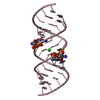
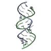
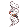
| Title | 1.8 A X-ray crystal structure of Apramycin complex with RNA fragment GGGCGUCGCUAGUACC/CGGUACUAAAAGUCGCC containing the human ribosomal decoding A site: RNA construct with 5'-overhang | ||||||
 Components Components |
| ||||||
 Keywords Keywords | RNA / Aminoglycoside antibiotics / Apramycin / Ribosomal decoding site / A site / Homo sapiens / RNA duplex | ||||||
| Function / homology | APRAMYCIN / RNA / RNA (> 10) Function and homology information Function and homology information | ||||||
| Method |  X-RAY DIFFRACTION / SYNCHROTRON / MOLECULAR REPLACEMENT / Resolution: 1.8 Å X-RAY DIFFRACTION / SYNCHROTRON / MOLECULAR REPLACEMENT / Resolution: 1.8 Å | ||||||
 Authors Authors | Hermann, T. / Tereshko, V. / Skripkin, E. / Patel, D.J. | ||||||
 Citation Citation | Journal: Blood Cells Mol.Dis. / Year: 2007 Title: Apramycin recognition by the human ribosomal decoding site. Authors: Hermann, T. / Tereshko, V. / Skripkin, E. / Patel, D.J. | ||||||
| History |
| ||||||
| Remark 999 | SEQUENCE THE RESIDUES THAT ARE NATURALLY OCCURING IN THE HUMAN RIBOSOMAL DECODING A SITE ARE ...SEQUENCE THE RESIDUES THAT ARE NATURALLY OCCURING IN THE HUMAN RIBOSOMAL DECODING A SITE ARE NUMBERED 1404-1413 (CHAIN A) AND 1487-1497 (CHAIN B). THIS NUMBERING IS ADAPTED FROM THE BACTERIAL ESCHERICHIA COLI SEQUENCE. IN BOTH STRANDS TERMINAL RESIDUES ARE DISTINCT FROM HUMAN AND WERE INCORPORATED FOR CRYSTALLIZATION PURPOSES. THE INCORPORATED RESIDUES ARE NUMBERED 1-3, 14-16 (CHAIN A) AND 83-86,98-99 (CHAIN B) |
- Structure visualization
| Structure viewer | Molecule: MolmilJmol/JSmol |
|---|
- Downloads & links
-Download
| PDBx/mmCIF format | 2oe8.cif.gz | 34.5 KB | Display | PDBx/mmCIF format |
|---|---|---|---|---|
| PDB format | pdb2oe8.ent.gz | 23.4 KB | Display | PDB format |
| PDBx/mmJSON format | 2oe8.json.gz | Tree view | PDBx/mmJSON format | |
| Others |  Other downloads Other downloads |
-Validation report
| Arichive directory | https://data.pdbj.org/pub/pdb/validation_reports/oe/2oe8ftp://data.pdbj.org/pub/pdb/validation_reports/oe/2oe8 | HTTPS FTP |
|---|
-Related structure data
| Related structure data | 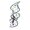 2oe5SC  2oe6C S: Starting model for refinement C: citing same article ( |
|---|---|
| Similar structure data |


-Links
 PDBj
PDBj


- Assembly
| Deposited unit | 
| ||||||||
|---|---|---|---|---|---|---|---|---|---|
| 1 |
| ||||||||
| Unit cell |
|
-Components
| #1: RNA chain | Mass: 5129.095 Da / Num. of mol.: 1 / Source method: obtained synthetically / Details: chemically synthesized |
|---|---|
| #2: RNA chain | Mass: 5426.301 Da / Num. of mol.: 1 / Source method: obtained synthetically / Details: chemically synthesized |
| #3: Chemical | ChemComp-AM2 /   Mass: 539.577 Da / Num. of mol.: 1 / Source method: obtained synthetically / Formula: C21H41N5O11 Mass: 539.577 Da / Num. of mol.: 1 / Source method: obtained synthetically / Formula: C21H41N5O11 |
| #4: Water | ChemComp-HOH /  Mass: 18.015 Da / Num. of mol.: 151 / Source method: isolated from a natural source / Formula: H2O Mass: 18.015 Da / Num. of mol.: 151 / Source method: isolated from a natural source / Formula: H2O |
-Experimental details
-Experiment
| Experiment | Method: X-RAY DIFFRACTION / Number of used crystals: 1 |
|---|
- Sample preparation
| Crystal | Density Matthews: 2.1 Å3/Da / Density % sol: 41.54 % | ||||||||||||||||||||||||
|---|---|---|---|---|---|---|---|---|---|---|---|---|---|---|---|---|---|---|---|---|---|---|---|---|---|
| Crystal grow | Temperature: 298 K / Method: vapor diffusion, hanging drop / pH: 5.6 Details: 0.05M MES BUFFER, 15-20% MPD, 0.02-0.04M MAGNESIUM ACETATE, pH 6, VAPOR DIFFUSION, HANGING DROP, temperature 298K, pH 5.6 | ||||||||||||||||||||||||
| Components of the solutions |
|
-Data collection
| Diffraction | Mean temperature: 100 K |
|---|---|
| Diffraction source | Source: SYNCHROTRON / Site: NSLS  / Beamline: X9A / Wavelength: 1 Å / Beamline: X9A / Wavelength: 1 Å |
| Detector | Type: ADSC QUANTUM 4 / Detector: CCD / Date: Apr 1, 2000 |
| Radiation | Protocol: SINGLE WAVELENGTH / Monochromatic (M) / Laue (L): M / Scattering type: x-ray |
| Radiation wavelength | Wavelength: 1 Å / Relative weight: 1 |
| Reflection | Resolution: 1.8→20 Å / Num. all: 8704 / Num. obs: 8704 / % possible obs: 99.44 % / Observed criterion σ(F): 0 / Observed criterion σ(I): 0 |
- Processing
| Software |
| ||||||||||||||||||||||||||||||||||||||||||||||||||||||||||||||||||||||||||||||||||||||||||||||||||||||||||||||||||||||||||||||||||||||||||||||||||||||||||||||||||||||||||
|---|---|---|---|---|---|---|---|---|---|---|---|---|---|---|---|---|---|---|---|---|---|---|---|---|---|---|---|---|---|---|---|---|---|---|---|---|---|---|---|---|---|---|---|---|---|---|---|---|---|---|---|---|---|---|---|---|---|---|---|---|---|---|---|---|---|---|---|---|---|---|---|---|---|---|---|---|---|---|---|---|---|---|---|---|---|---|---|---|---|---|---|---|---|---|---|---|---|---|---|---|---|---|---|---|---|---|---|---|---|---|---|---|---|---|---|---|---|---|---|---|---|---|---|---|---|---|---|---|---|---|---|---|---|---|---|---|---|---|---|---|---|---|---|---|---|---|---|---|---|---|---|---|---|---|---|---|---|---|---|---|---|---|---|---|---|---|---|---|---|---|---|
| Refinement | Method to determine structure: MOLECULAR REPLACEMENT Starting model: 2OE5 Resolution: 1.8→20 Å / Cor.coef. Fo:Fc: 0.963 / Cor.coef. Fo:Fc free: 0.952 / SU B: 3.144 / SU ML: 0.094 / TLS residual ADP flag: LIKELY RESIDUAL / Cross valid method: THROUGHOUT / ESU R: 0.166 / ESU R Free: 0.142 / Stereochemistry target values: MAXIMUM LIKELIHOOD / Details: HYDROGENS HAVE BEEN ADDED IN THE RIDING POSITIONS
| ||||||||||||||||||||||||||||||||||||||||||||||||||||||||||||||||||||||||||||||||||||||||||||||||||||||||||||||||||||||||||||||||||||||||||||||||||||||||||||||||||||||||||
| Solvent computation | Ion probe radii: 0.8 Å / Shrinkage radii: 0.8 Å / VDW probe radii: 1.4 Å / Solvent model: BABINET MODEL WITH MASK | ||||||||||||||||||||||||||||||||||||||||||||||||||||||||||||||||||||||||||||||||||||||||||||||||||||||||||||||||||||||||||||||||||||||||||||||||||||||||||||||||||||||||||
| Displacement parameters | Biso mean: 24.678 Å2
| ||||||||||||||||||||||||||||||||||||||||||||||||||||||||||||||||||||||||||||||||||||||||||||||||||||||||||||||||||||||||||||||||||||||||||||||||||||||||||||||||||||||||||
| Refinement step | Cycle: LAST / Resolution: 1.8→20 Å
| ||||||||||||||||||||||||||||||||||||||||||||||||||||||||||||||||||||||||||||||||||||||||||||||||||||||||||||||||||||||||||||||||||||||||||||||||||||||||||||||||||||||||||
| Refine LS restraints |
| ||||||||||||||||||||||||||||||||||||||||||||||||||||||||||||||||||||||||||||||||||||||||||||||||||||||||||||||||||||||||||||||||||||||||||||||||||||||||||||||||||||||||||
| LS refinement shell | Resolution: 1.8→1.846 Å / Total num. of bins used: 20 /
| ||||||||||||||||||||||||||||||||||||||||||||||||||||||||||||||||||||||||||||||||||||||||||||||||||||||||||||||||||||||||||||||||||||||||||||||||||||||||||||||||||||||||||
| Refinement TLS params. | Method: refined / Origin x: 18.2279 Å / Origin y: -1.6955 Å / Origin z: 42.5781 Å
| ||||||||||||||||||||||||||||||||||||||||||||||||||||||||||||||||||||||||||||||||||||||||||||||||||||||||||||||||||||||||||||||||||||||||||||||||||||||||||||||||||||||||||
| Refinement TLS group |
|
