 Movie
Movie Controller

[English] 日本語
 Yorodumi
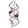
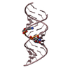
- PDB-1z7f: Crystal structure of 16 base pair RNA duplex containing a C-A mismatch -
+ Open data

- Basic information
| Entry | Database: PDB / ID: 1z7f | ||||||||||||||||||
|---|---|---|---|---|---|---|---|---|---|---|---|---|---|---|---|---|---|---|---|
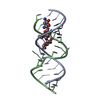
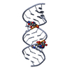
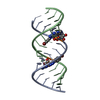
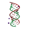
| Title | Crystal structure of 16 base pair RNA duplex containing a C-A mismatch | ||||||||||||||||||
 Components Components | 5'-R(* Keywords KeywordsRNA / DUPLEX / MISMATCH | Function / homology | STRONTIUM ION / RNA / RNA (> 10) |  Function and homology information Function and homology informationMethod |  X-RAY DIFFRACTION / MOLECULAR REPLACEMENT / Resolution: 2.1 Å X-RAY DIFFRACTION / MOLECULAR REPLACEMENT / Resolution: 2.1 Å  Authors AuthorsGherghe, C.M. / Krahn, J.M. / Weeks, K.M. |  CitationJournal: J.Am.Chem.Soc. / Year: 2005 CitationJournal: J.Am.Chem.Soc. / Year: 2005Title: Crystal structures, reactivity and inferred acylation transition States for 2'-amine substituted RNA. Authors: Gherghe, C.M. / Krahn, J.M. / Weeks, K.M. History |
|
- Structure visualization
| Structure viewer | Molecule: MolmilJmol/JSmol |
|---|
- Downloads & links
-Download
| PDBx/mmCIF format | 1z7f.cif.gz | 38.9 KB | Display | PDBx/mmCIF format |
|---|---|---|---|---|
| PDB format | pdb1z7f.ent.gz | 25.8 KB | Display | PDB format |
| PDBx/mmJSON format | 1z7f.json.gz | Tree view | PDBx/mmJSON format | |
| Others |  Other downloads Other downloads |
-Validation report
| Arichive directory | https://data.pdbj.org/pub/pdb/validation_reports/z7/1z7fftp://data.pdbj.org/pub/pdb/validation_reports/z7/1z7f | HTTPS FTP |
|---|
-Related structure data
| Related structure data | 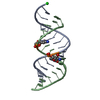 1yrmSC 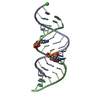 1yy0C  1yzdC  1z79C S: Starting model for refinement C: citing same article ( |
|---|---|
| Similar structure data | |
| Other databases |
|


-Links
 PDBj
PDBj


- Assembly
| Deposited unit | 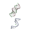
| ||||||||
|---|---|---|---|---|---|---|---|---|---|
| 1 | 
| ||||||||
| 2 | 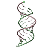
| ||||||||
| Unit cell |
| ||||||||
| Components on special symmetry positions |
|
-Components
| #1: RNA chain | Mass: 5082.079 Da / Num. of mol.: 3 / Source method: obtained synthetically #2: Chemical |   Mass: 87.620 Da / Num. of mol.: 2 / Source method: obtained synthetically / Formula: Sr Mass: 87.620 Da / Num. of mol.: 2 / Source method: obtained synthetically / Formula: Sr#3: Water | ChemComp-HOH / |  Mass: 18.015 Da / Num. of mol.: 67 / Source method: isolated from a natural source / Formula: H2O Mass: 18.015 Da / Num. of mol.: 67 / Source method: isolated from a natural source / Formula: H2O |
|---|
-Experimental details
-Experiment
| Experiment | Method: X-RAY DIFFRACTION / Number of used crystals: 1 |
|---|
- Sample preparation
| Crystal | Density Matthews: 2.14 Å3/Da / Density % sol: 42.4 % | ||||||||||||||||||||||||||||||||||||||||||||
|---|---|---|---|---|---|---|---|---|---|---|---|---|---|---|---|---|---|---|---|---|---|---|---|---|---|---|---|---|---|---|---|---|---|---|---|---|---|---|---|---|---|---|---|---|---|
| Crystal grow | Temperature: 273 K / Method: vapor diffusion, sitting drop / pH: 7 Details: MPD, sodium cacodylate, spermine, strontium chloride, magnesium chloride, pH 7.0, VAPOR DIFFUSION, SITTING DROP, temperature 273K | ||||||||||||||||||||||||||||||||||||||||||||
| Components of the solutions |
|
-Data collection
| Diffraction | Mean temperature: 100 K |
|---|---|
| Diffraction source | Source: ROTATING ANODE / Type: RIGAKU / Wavelength: 1.5418 Å |
| Detector | Type: RIGAKU / Detector: IMAGE PLATE / Date: Feb 14, 2004 |
| Radiation | Protocol: SINGLE WAVELENGTH / Monochromatic (M) / Laue (L): M / Scattering type: x-ray |
| Radiation wavelength | Wavelength: 1.5418 Å / Relative weight: 1 |
| Reflection | Resolution: 2→27 Å / Num. all: 9573 / Num. obs: 8972 / % possible obs: 93.8 % / Observed criterion σ(I): 1 / Redundancy: 12.02 % / Biso Wilson estimate: 26.5 Å2 / Rmerge(I) obs: 0.083 / Rsym value: 0.108 / Net I/σ(I): 22.11 |
| Reflection shell | Resolution: 2→2.07 Å / Rmerge(I) obs: 0.176 / Mean I/σ(I) obs: 4.59 / Num. unique all: 638 / Rsym value: 0.158 / % possible all: 68.7 |
- Processing
| Software |
| ||||||||||||||||||||||||||||||||||||
|---|---|---|---|---|---|---|---|---|---|---|---|---|---|---|---|---|---|---|---|---|---|---|---|---|---|---|---|---|---|---|---|---|---|---|---|---|---|
| Refinement | Method to determine structure: MOLECULAR REPLACEMENT Starting model: pdb entry 1YRM Resolution: 2.1→19.01 Å / Rfactor Rfree error: 0.013 / Data cutoff high absF: 1006060.31 / Data cutoff low absF: 0 / Isotropic thermal model: RESTRAINED / Cross valid method: THROUGHOUT / σ(F): 0
| ||||||||||||||||||||||||||||||||||||
| Solvent computation | Solvent model: FLAT MODEL / Bsol: 44.0469 Å2 / ksol: 0.353048 e/Å3 | ||||||||||||||||||||||||||||||||||||
| Displacement parameters | Biso mean: 33.4 Å2
| ||||||||||||||||||||||||||||||||||||
| Refine analyze |
| ||||||||||||||||||||||||||||||||||||
| Refinement step | Cycle: LAST / Resolution: 2.1→19.01 Å
| ||||||||||||||||||||||||||||||||||||
| Refine LS restraints |
| ||||||||||||||||||||||||||||||||||||
| LS refinement shell | Resolution: 2.1→2.23 Å / Rfactor Rfree error: 0.039 / Total num. of bins used: 6
| ||||||||||||||||||||||||||||||||||||
| Xplor file |
|
