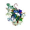
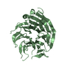
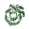
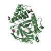
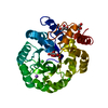
- PDB-2k2f: Solution structure of Ca2+-S100A1-RyRP12 -
+
データを開く
IDまたはキーワード:
読み込み中...
-
基本情報
登録情報
データベース: PDB / ID: 2k2f
タイトル
Solution structure of Ca2+-S100A1-RyRP12
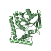
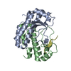
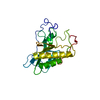
要素
Protein S100-A1
Ryanodine receptor 1 peptide
キーワード
METAL BINDING PROTEIN / S100 / EF hand / ryanodine receptor / calcium binding / Alternative splicing / Calcium channel / Calcium transport / Glycoprotein / Ion transport / Ionic channel / Membrane / Polymorphism / Transmembrane / Transport / Cytoplasm / Metal-binding / Zinc
機能・相同性
機能・相同性情報
manganese ion transmembrane transport / Stimuli-sensing channels / Regulation of TLR by endogenous ligand / sarcoplasmic reticulum calcium ion transport / establishment of protein localization to endoplasmic reticulum / type B pancreatic cell apoptotic process / Purkinje myocyte to ventricular cardiac muscle cell signaling / A band / calcium-induced calcium release activity / regulation of atrial cardiac muscle cell action potential ...manganese ion transmembrane transport / Stimuli-sensing channels / Regulation of TLR by endogenous ligand / sarcoplasmic reticulum calcium ion transport / establishment of protein localization to endoplasmic reticulum / type B pancreatic cell apoptotic process / Purkinje myocyte to ventricular cardiac muscle cell signaling / A band / calcium-induced calcium release activity / regulation of atrial cardiac muscle cell action potential / left ventricular cardiac muscle tissue morphogenesis / suramin binding / regulation of AV node cell action potential / regulation of SA node cell action potential / regulation of ventricular cardiac muscle cell action potential / ventricular cardiac muscle cell action potential / positive regulation of sequestering of calcium ion / embryonic heart tube morphogenesis / cardiac muscle hypertrophy / S100 protein binding / calcium ion transport into cytosol / M band / ryanodine-sensitive calcium-release channel activity / I band / response to caffeine / regulation of cardiac muscle contraction by calcium ion signaling / release of sequestered calcium ion into cytosol by sarcoplasmic reticulum / extrinsic component of cytoplasmic side of plasma membrane / response to redox state / cellular response to caffeine / Ion homeostasis / calcium ion transmembrane import into cytosol / negative regulation of cytosolic calcium ion concentration / regulation of heart contraction / response to muscle activity / protein kinase A regulatory subunit binding / positive regulation of sprouting angiogenesis / protein kinase A catalytic subunit binding / positive regulation of the force of heart contraction / smooth endoplasmic reticulum / intracellularly gated calcium channel activity / response to magnesium ion / detection of calcium ion / regulation of cardiac muscle contraction / regulation of cytosolic calcium ion concentration / regulation of cardiac muscle contraction by regulation of the release of sequestered calcium ion / positive regulation of heart rate / cardiac muscle contraction / response to muscle stretch / release of sequestered calcium ion into cytosol / response to nutrient / cellular response to epinephrine stimulus / calcium channel complex / sarcoplasmic reticulum membrane / regulation of heart rate / sarcoplasmic reticulum / sarcomere / establishment of localization in cell / calcium-mediated signaling / response to calcium ion / sarcolemma / calcium ion transmembrane transport / calcium channel activity / Z disc / intracellular calcium ion homeostasis / calcium-dependent protein binding / calcium ion transport / nuclear envelope / ATPase binding / scaffold protein binding / monoatomic ion transmembrane transport / calmodulin binding / response to hypoxia / response to xenobiotic stimulus / calcium ion binding / protein kinase binding / enzyme binding / negative regulation of transcription by RNA polymerase II / Golgi apparatus / protein homodimerization activity / protein-containing complex / mitochondrion / nucleoplasm / identical protein binding / nucleus / membrane / cytosol / cytoplasm 類似検索 - 分子機能
Protein S100-A1 / S-100/ICaBP type calcium binding protein signature. / S100/Calcium binding protein 7/8-like, conserved site / S100/CaBP-9k-type, calcium binding, subdomain / S-100/ICaBP type calcium binding domain / S-100/ICaBP type calcium binding domain / EF hand domain / Ryanodine receptor, SPRY domain 2 / : / Ryanodine receptor junctional solenoid repeat ...Protein S100-A1 / S-100/ICaBP type calcium binding protein signature. / S100/Calcium binding protein 7/8-like, conserved site / S100/CaBP-9k-type, calcium binding, subdomain / S-100/ICaBP type calcium binding domain / S-100/ICaBP type calcium binding domain / EF hand domain / Ryanodine receptor, SPRY domain 2 / : / Ryanodine receptor junctional solenoid repeat / Ryanodine Receptor TM 4-6 / Ryanodine receptor / Ryanodine receptor, SPRY domain 1 / Ryanodine receptor, SPRY domain 3 / Ryanodine Receptor TM 4-6 / Ryanodine receptor Ryr / RyR domain / RyR/IP3 receptor binding core, RIH domain superfamily / RyR/IP3R Homology associated domain / Inositol 1,4,5-trisphosphate/ryanodine receptor / RIH domain / RyR and IP3R Homology associated / Inositol 1,4,5-trisphosphate/ryanodine receptor / RIH domain / : / MIR motif / MIR domain / MIR domain profile. / Domain in ryanodine and inositol trisphosphate receptors and protein O-mannosyltransferases / Mir domain superfamily / SPRY domain / B30.2/SPRY domain / B30.2/SPRY domain profile. / B30.2/SPRY domain superfamily / Domain in SPla and the RYanodine Receptor. / SPRY domain / EF-hand / Recoverin; domain 1 / EF-hand domain pair / EF-hand, calcium binding motif / EF-Hand 1, calcium-binding site / EF-hand calcium-binding domain. / EF-hand calcium-binding domain profile. / EF-hand domain / Ion transport domain / Ion transport protein / EF-hand domain pair / Concanavalin A-like lectin/glucanase domain superfamily / Orthogonal Bundle / Mainly Alpha 類似検索 - ドメイン・相同性
Ryanodine receptor 2 / Protein S100-A1 類似検索 - 構成要素
 ムービー
ムービー コントローラー


 データを開く
 基本情報
基本情報 要素
要素 キーワード
キーワード 機能・相同性情報
 データ登録者
 引用
引用 構造の表示
構造の表示 ダウンロードとリンク
 その他のダウンロード
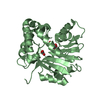

 PDBj
PDBj


 集合体
集合体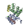


 分子量: 40.078 Da / 分子数: 4 / 由来タイプ: 合成 / 式: Ca
 HSQC
HSQC 試料調製
試料調製 解析
解析